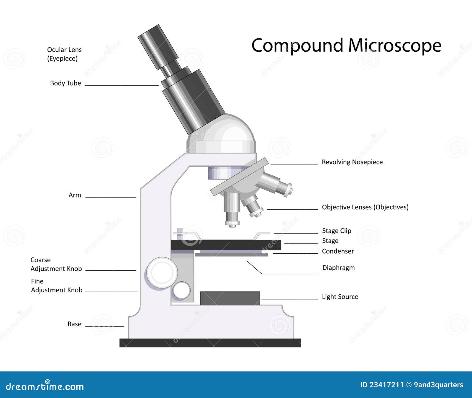
43 labeled diagram of compound microscope
Compound Microscope – Diagram (Parts labelled), Principle and ... Feb 03, 2022 · See: Labeled Diagram showing differences between compound and simple microscope parts Structural Components. The three structural components include. 1. Head. This is the upper part of the microscope that houses the optical parts. 2. Arm . This part connects the head with the base and provides stability to the microscope. Compound Microscope Labeled Diagram | Quizlet QUESTION. The total magnification of a specimen being viewed with a 10X ocular lens and a 40X objective lens is. 15 answers. QUESTION. a mosquito beats its wings up and down 600 times per second, which you hear as a very annoying 600 Hz sound. if the air outside is 20 C, how far would a sound wave travel between wing beats. 2 answers.
Microscope Parts, Function, & Labeled Diagram - slidingmotion Microscope parts labeled diagram gives us all the information about its parts and their position in the microscope. Microscope Parts Labeled Diagram, The principle of the Microscope gives you an exact reason to use it. It works on the 3 principles. Magnification, Resolving Power, Numerical Aperture. Parts of Microscope, Head, Base, Arm,

Labeled diagram of compound microscope
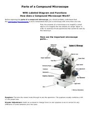
Compound Microscope Parts, Functions, and Labeled Diagram The total magnification of a compound microscope is calculated by multiplying the objective lens magnification by the eyepiece magnification level. So, a compound microscope with a 10x eyepiece magnification looking through the 40x objective lens has a total magnification of 400x (10 x 40). Solved Label the image of a compound light microscope using - Chegg Expert Answer. 100% (17 ratings) Transcribed image text: Label the image of a compound light microscope using the terms provided. Microscope Parts and Functions With Labeled Diagram and ... Before exploring microscope parts and functions, you should probably understand that the compound light microscope is more complicated than just a microscope with more than one lens. First, the purpose of a microscope is to magnify a small object or to magnify the fine details of a larger object in order to examine minute specimens that cannot ...
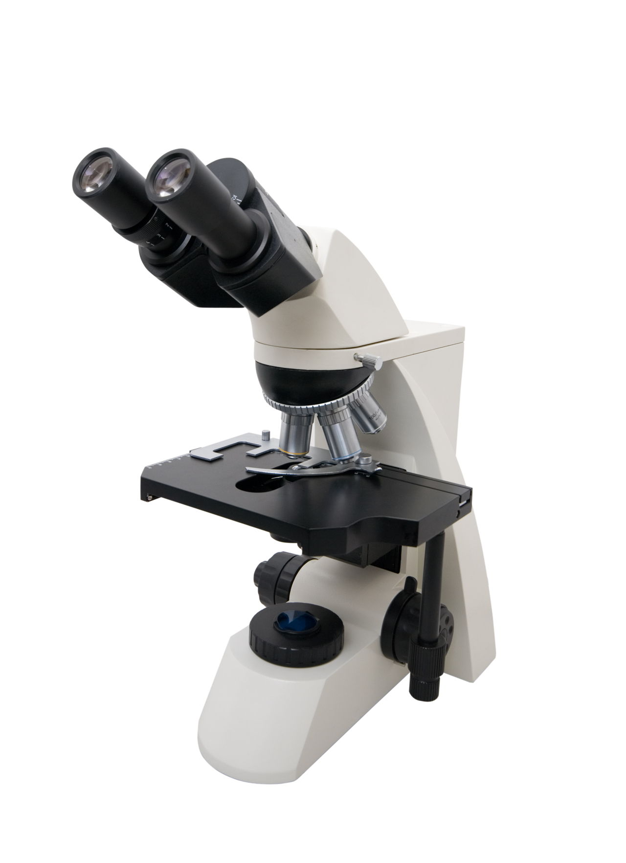
Labeled diagram of compound microscope. Compound Microscope- Definition, Labeled Diagram, Principle ... Apr 03, 2022 · Parts of a Compound Microscope. Eyepiece And Body Tube. The eyepiece is the lens through which the viewer looks to see the specimen. It usually contains a 10X or 15X power lens. The body tube connects the eyepiece to the objective lenses. Objectives and Stage Clips. Objective Lenses are one of the most important parts of a Compound Microscope. (i) Draw a neat labelled ray diagram of a compound microscope. Explain ... The eyepiece forms its image A'' B'' which is virtual, erect and magnified. Thus the final image A'' B'' formed by the microscope is inverted and magnified and its position is outside the objective and eyepiece towards objective lens. Magnifying power of compound microscope is. for final image at distance of distinct vision. for final image at ... 16 Parts of a Compound Microscope: Diagrams and Video In compound microscopes with two eye pieces there are prisms contained in the body that will also split the beam of light to enable you to view the image through both eye pieces. 2. Arm. The arm of the microscope is another structural piece. The arm connects the base of the microscope to the head/body of the microscope. Compound Microscope - Types, Parts, Diagram, Functions and Uses It comes with a wide body and base. Its distinct parts include a condenser, illumination, focus lock, mechanical stage, and a revolving nosepiece which can hold up to five objectives. It usually has a binocular head, which makes long-term observation easy. Image 22: An example of a research compound microscope.
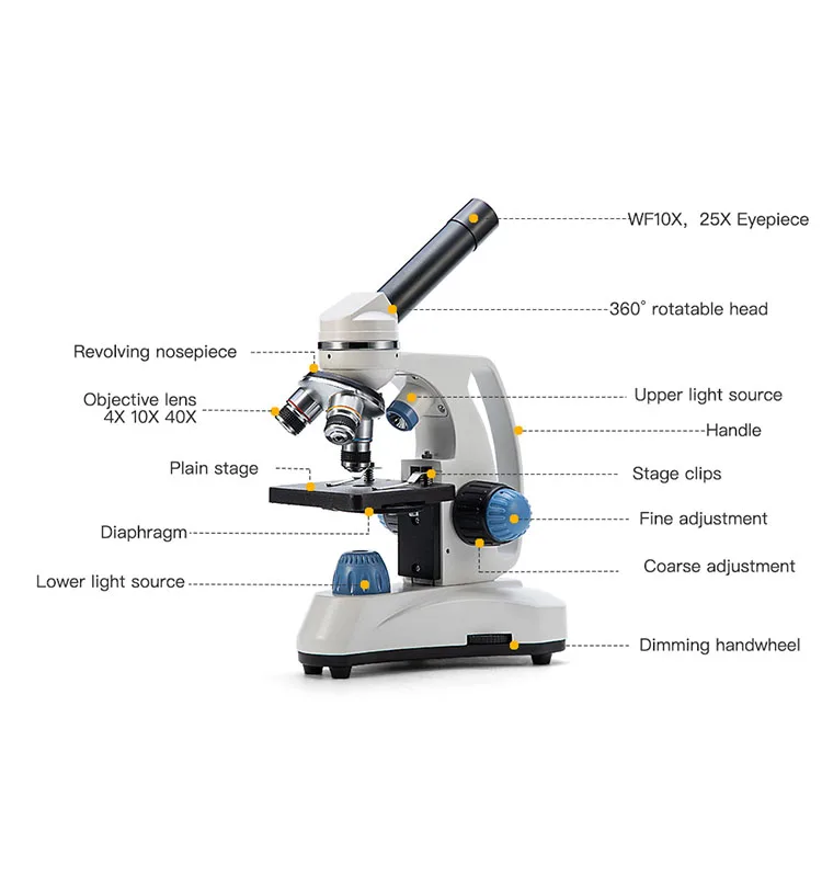
Microscope, Microscope Parts, Labeled Diagram, and Functions Sep 03, 2022 · Simply multiply the magnification of the ocular lens by the magnification of the objective lens to calculate the power of magnification of a microscope. For a typical compound microscope with a 10X ocular lens and objective lenses with magnifications of 4X, 10X, 40X, and 100X, your microscope will have magnifications of 40X, 100X, 400X, and ... Label the microscope — Science Learning Hub Use this interactive to identify and label the main parts of a microscope. Drag and drop the text labels onto the microscope diagram. eye piece lens, diaphragm or iris, coarse focus adjustment, stage, base, fine focus adjustment, light source, high-power objective, Download Exercise, Tweet, A Study of the Microscope and its Functions With a Labeled Diagram ... These labeled microscope diagrams and the functions of its various parts, attempt to simplify the microscope for you. However, as the saying goes, 'practice makes perfect', here is a blank compound microscope diagram and blank electron microscope diagram to label. Download the diagrams and practice labeling the different parts of these ... Compound Microscope Parts, Function, & Diagram | What is a Compound ... The base of the compound light microscope is the bottom portion of the compound microscope. It functions to support the entire compound microscope. The base can be set on a table or lab bench, and...
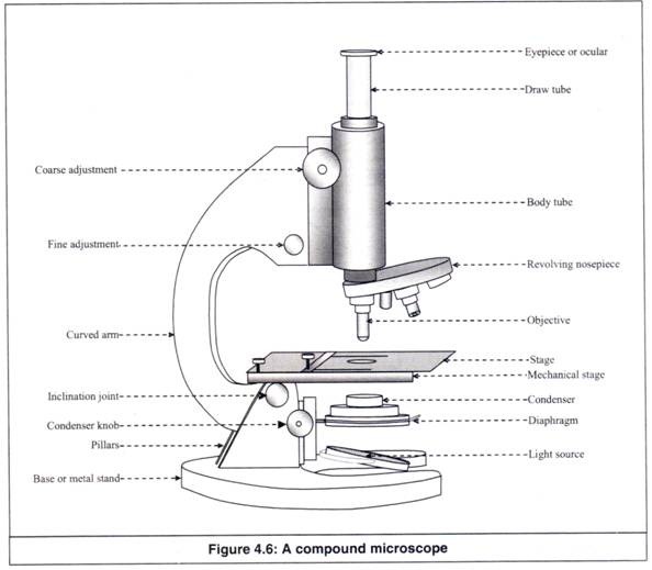
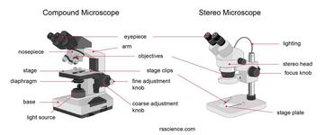
Compound Microscope: Parts of Compound Microscope - BYJUS (A) Mechanical Parts of a Compound Microscope, 1. Foot or base, It is a U-shaped structure and supports the entire weight of the compound microscope. 2. Pillar, It is a vertical projection. This stands by resting on the base and supports the stage. 3. Arm, The entire microscope is handled by a strong and curved structure known as the arm. 4. Stage, Electron Microscope Principle, Uses, Types and Images ... Feb 02, 2022 · Ans: A light microscope has a low resolving power (0.25µm to 0.3µm) while the electron microscope has a resolution power about 250 times higher than the light microscope at about 0.001µm. Similarly, a light microscope has a magnification of 500X to 1500x while the electron microscope has a much higher magnification of 100,000X to 300,000X. Parts of Stereo Microscope (Dissecting microscope) – labeled ... If you would like to learn optical components of a compound microscope, please visit Compound Microscope Parts – Labeled Diagram and their Functions, and this article. How to use a stereo (dissecting) microscope. Follow these steps to put your stereo microscopes in work: 1. Set your microscope on a tabletop or other flat sturdy surface where ... Microscope Types (with labeled diagrams) and Functions Compound microscope labeled diagram, Compound microscope functions: It finds great application in areas of pathology, pedology, forensics etc, Its greater order of magnification allows for deeper study of microbial organisms to, Detect the cause of diseases, Study the mineral composition in soils,
Biology 4 U on Twitter: "Try this labelled diagram Quiz on ...
parts of a microscope diagram Microscope parts science compound knob adjustment coarse labeled diagram biology microscopes light label name tools lab functions does structure 6th. Plant phloem vascular cells anatomy parenchyma primary cambium sclereids fibers tissue sieve elements conducting differentiated procambium initiated secondary called grkraj,

Labeled Microscope Diagram | Microscope parts, Science fair ...
Label a Compound Microscope Diagram | Quizlet Start studying Label a Compound Microscope. Learn vocabulary, terms, and more with flashcards, games, and other study tools.

Compound Microscope: Parts of Compound Microscope
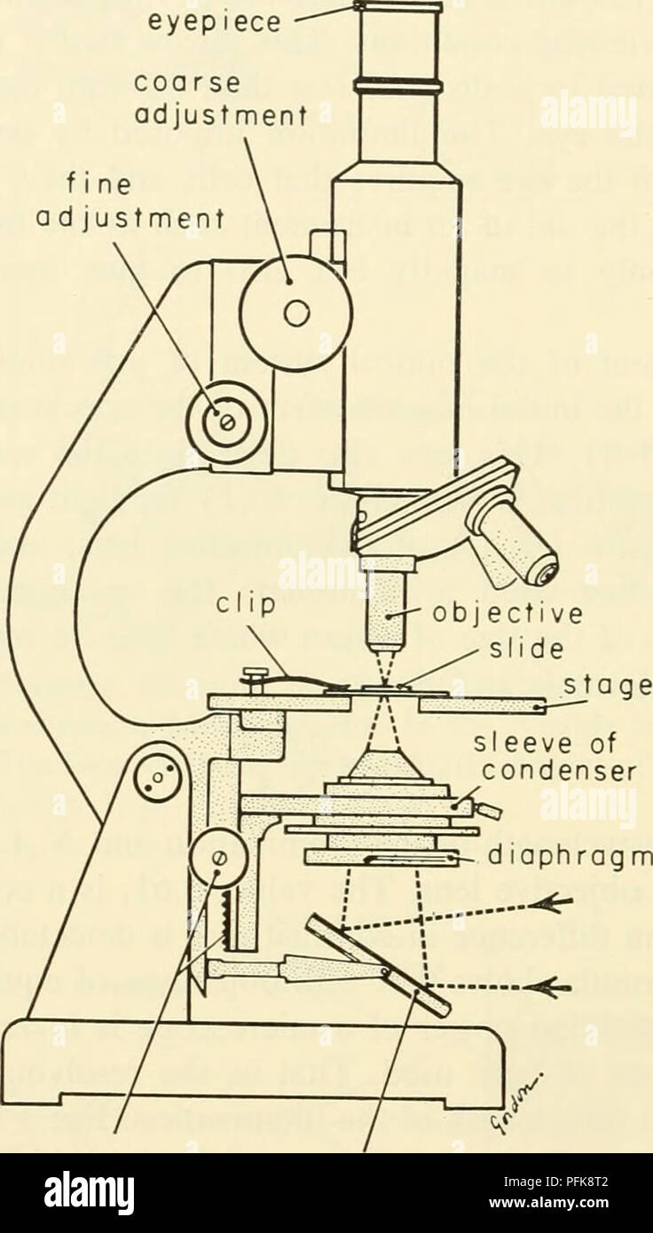
Parts of a microscope with functions and labeled diagram Apr 19, 2022 · Figure: Diagram of parts of a microscope. There are three structural parts of the microscope i.e. head, base, and arm. Head – This is also known as the body. It carries the optical parts in the upper part of the microscope. Base – It acts as microscopes support. It also carries microscopic illuminators.

Labeling the Parts of the Microscope | Microscope World Resources
Diagram of a Compound Microscope - Biology Discussion The size of objects viewed under the compound microscope can be accurately determined using a micrometer. The latter consists of two scales, the eyepiece scale, (also called 'graticule' or 'ocular') and the stage micrometer scale. The eyepiece scale is calibrated with the help of stage micrometer and the former is then used for measurements.

Parts of a microscope with functions and labeled diagram
Solved Part III. Label the following diagram of a compound | Chegg.com Label the following diagram of a compound light microscope Match each of the following parts of the microscope to the correct description. Use the descriptions as often as needed. - Stage a) Series of tubes that house the second lens Objective Lenses system: scanning, low.power, high-power, and oil immersion. Ocular Lens b) A flat platform where

Microscope Labeling Diagram | Quizlet
Compound Microscope Parts - Labeled Diagram and their Functions Labeled diagram of a compound microscope, Major structural parts of a compound microscope, Optical components of a compound microscope, Eyepiece, Eyepiece tube, Objective lenses, Nosepiece, Specimen stage, Coarse and fine focus knobs, Rack stop, Illuminator, Condenser, Abbe condenser, Iris Diaphragm, Condenser Focus Knob, Summary,

Compound Microscope Parts – Labeled Diagram and their ...
Compound Microscope: Definition, Diagram, Parts, Uses, Working ... - BYJUS The parts of a compound microscope can be classified into two: Non-optical parts, Optical parts, Non-optical parts, Base, The base is also known as the foot which is either U or horseshoe-shaped. It is a metallic structure that supports the entire microscope. Pillar, The connection between the base and the arm are possible through the pillar. Arm,

Compound Microscope Parts – Labeled Diagram and their ...
Binocular Microscope Anatomy - Parts and Functions with a Labeled Diagram First, see the body and arm of the light compound microscope. The body tube is a cylindrical-like structure that connects the ocular lens to the objective lenses. Again, the arm of the microscope connects the body tube to the microscope's base. You will see the coarse and fine adjustment in the arm of the microscope.

File:Labelledmicroscope.gif - Wikimedia Commons
Parts of a Compound Microscope and Their Functions - NotesHippo Compound microscope mechanical parts (Microscope Diagram: 2) include base or foot, pillar, arm, inclination joint, stage, clips, diaphragm, body tube, nose piece, coarse adjustment knob and fine adjustment knob. Base: It's the horseshoe-shaped base structure of microscope. All of the other components of the compound microscope are supported ...

File:Microscope diagram.png - Wikimedia Commons
(b) Why both objective and eyepiece of a compound microscope must have ... Question, (a) Draw the labelled ray diagram for the formation of image by a compound microscope. Derive an expression for its total magnification (or magnifying power), when the final image is formed at the near point. (b) Why both objective and eyepiece of a compound microscope must have short focal lengths?

Parts of Stereo Microscope (Dissecting microscope) – labeled ...
Draw a neat labelled diagram of a compound microscope and ... - Sarthaks Using sign convention, we find that O'I 1 = + v 0 and O'O = -u where v 0 is the image distance due to the objective and u is the object distance for the objective or the compound microscope. I 1 G 1 is negative and OJ is positive. To find me : The eyepiece behaves like a simple microscope. So : the magnifying power of the eye piece. ∴ m e ...

Compound Light Microscope Labeling Diagram | Quizlet
Labelled Diagram of Compound Microscope The below mentioned article provides a labelled diagram of compound microscope. Part # 1. The Stand: The stand is made up of a heavy foot which carries a curved inclinable limb or arm bearing the body tube. The foot is generally horse shoe-shaped structure (Fig. 2) which rests on table top or any other surface on which the microscope in kept.
parts of microscope with diagram - Clip Art Library
Microscope Parts and Functions With Labeled Diagram and ... Before exploring microscope parts and functions, you should probably understand that the compound light microscope is more complicated than just a microscope with more than one lens. First, the purpose of a microscope is to magnify a small object or to magnify the fine details of a larger object in order to examine minute specimens that cannot ...
Mikroskop Swift,Mikroskop Monokuler 40 X-1000x Untuk Anak Siswa Optik Biologi - Buy Mikroskop,Biological Microscope,Microscopio Product on Alibaba.com
Solved Label the image of a compound light microscope using - Chegg Expert Answer. 100% (17 ratings) Transcribed image text: Label the image of a compound light microscope using the terms provided.

Labeling the Parts of the Microscope | Microscope World Resources
Compound Microscope Parts, Functions, and Labeled Diagram The total magnification of a compound microscope is calculated by multiplying the objective lens magnification by the eyepiece magnification level. So, a compound microscope with a 10x eyepiece magnification looking through the 40x objective lens has a total magnification of 400x (10 x 40).

Compound Microscope Parts – Labeled Diagram and their ...

Microscope Diagram Labeled, Unlabeled and Blank | Parts of a ...

Microscope Diagram Labeled, Unlabeled and Blank | Parts of a ...
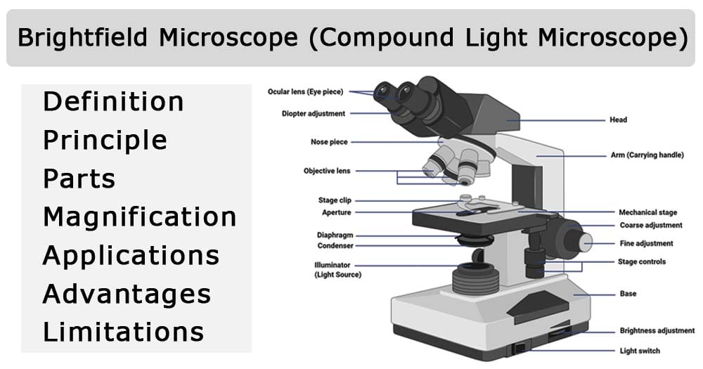
Brightfield Microscope (Compound Light Microscope ...

Compound Microscope Parts – Labeled Diagram and their ...

Diagram of a Compound Microscope
Parts of Stereo Microscope (Dissecting microscope) – labeled ...

label microscope diagram | Charts | Microscope, Anatomy bones ...
Diagram of a Microscope - Guide to using a microscope

Difference between Simple and Compound Microscope ...
Understanding the Compound Microscope Parts and its Functions ...
MICROBIO 16 Parts of a Compound Microscope with Diagram and ...
Cytology. Cytology. radiation used to illuminate the specimen ...
a) Draw a labelled ray diagram of a compound microscope. (b ...

This is a common compound microscope Label its parts class 11 ...
Compound Microscope stock vector. Illustration of research ...

Parts of Microscope, Function, Names & Labeled Diagram ...

Draw a well labelled diagram of a microscope. - Brainly.in

compound microscope Diagram | Quizlet

Microscope, Microscope Parts, Labeled Diagram, and Functions

Microscope diagram labeled | Clipart Panda - Free Clipart Images

This is a common compound microscope. Label its parts from A ...

Solved A. OLYMPUS C. B. Use the Diagram to answer the | Chegg.com

Draw a labelled diagram of an image formed by a compound ...

Parts of a Compound Microscope and Their Functions

Compound Microscope- Definition, Labeled Diagram, Principle ...

labelled diagram of microscope - Brainly.in

Parts of a microscope with functions and labeled diagram
Post a Comment for "43 labeled diagram of compound microscope"