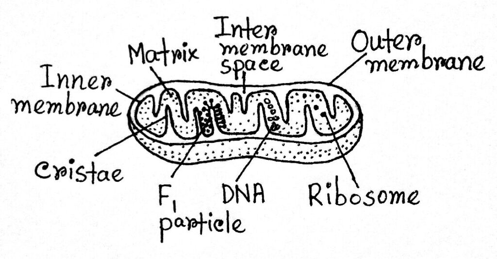
40 labelled diagram of mitochondria
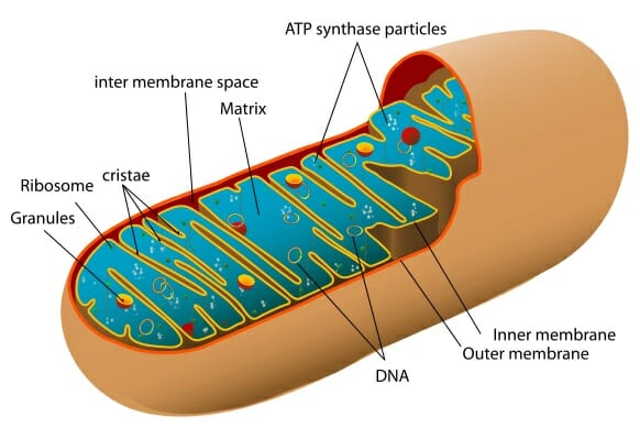
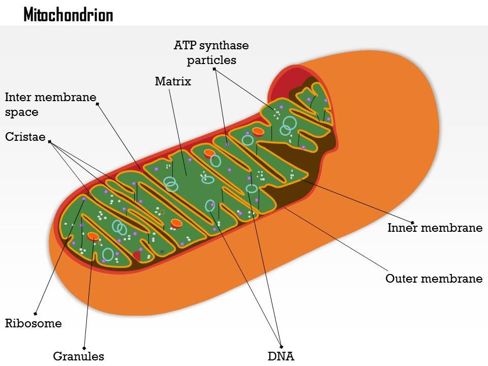
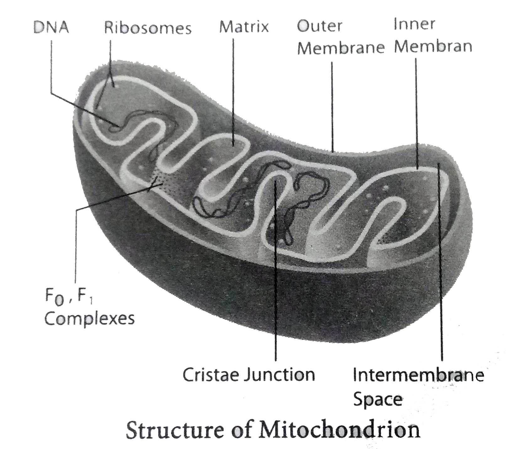
Mitochondria (video) | Khan Academy Mitochondria are organelles that contain their own DNA, and have both inner and outer membranes. Mitochondria are often referred to as the "powerhouses of the cell" because they are responsible for producing most of the cell's energy in the form of ATP. What Is Mitochondria (Structure, Diagram & Function) The diagram of mitochondria below illustrates several structural features of mitochondria. Structure of Mitochondria The mitochondrion is a double-membraned, rod-shaped structure found in both plant and animal cell. Its size ranges from 0.5 to 1.0 micrometre in diameter.
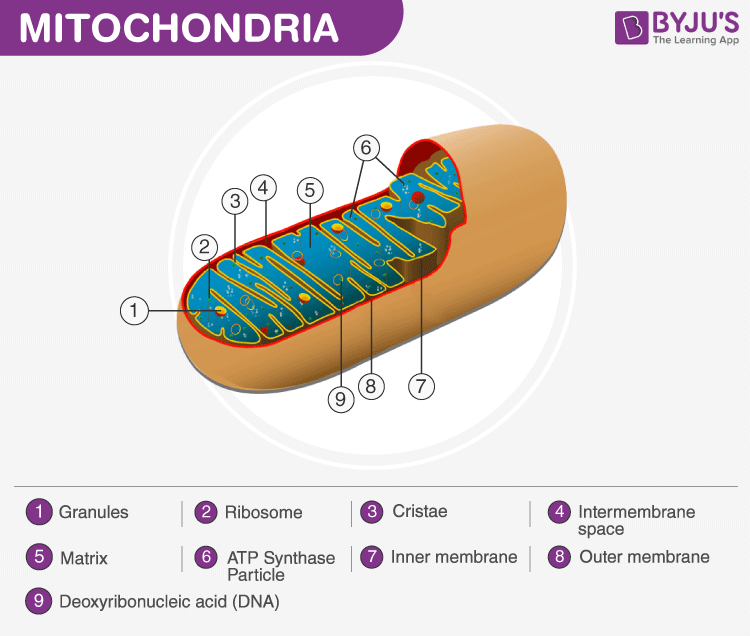
Diagram of Mitochondria | Labelled Diagram of Mitochondria A well-labelled diagram of mitochondria is given below for your better understanding of the structure. Labelled Diagram of a Mitochondrion Image will be updates soon Characteristics of the Mitochondrial DNA/ Genome: The mitochondrial DNA is circular and is made up of 16,569 DNA base pairs.

Labelled diagram of mitochondria
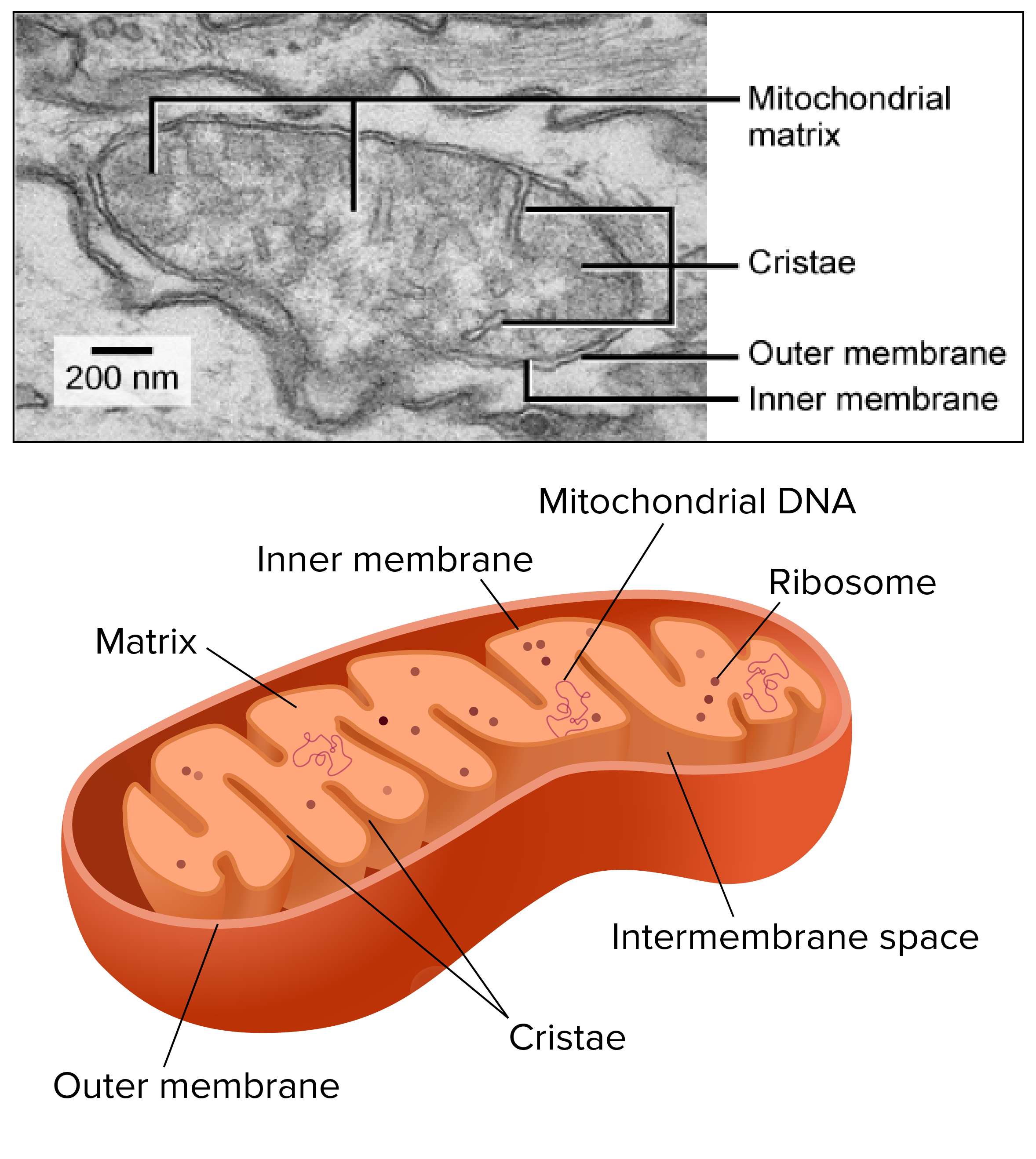
Mitochondria and chloroplasts The mitochondria are suspended in the jelly-like cytosol of the cell. They are oval-shaped and have two membranes: an outer one, surrounding the whole organelle, and an inner one, with many inward protrusions called cristae that increase surface area. Plant vs animal cells review A plant cell contains a large, singular vacuole that is used for storage and maintaining the shape of the cell. In contrast, animal cells have many, smaller vacuoles. Plant cells have a cell wall, as well as a cell membrane. In plants, the cell wall surrounds the cell membrane. This gives the plant cell its unique rectangular shape. Plant cells - Cell structure - AQA - GCSE Combined Science Revision ... A tiny organelle where protein synthesis occurs. Plant cells also have additional structures: Function. Chloroplast. Organelles that contains the green pigment, chlorophyll, which absorbs light ...
Labelled diagram of mitochondria. Mitochondria: Definition, Structure & Function (with Diagram) Mitochondria are membrane-bound organelles enclosed by a double membrane. They have a smooth outer membrane enclosing the organelle and a folded inner membrane. The folds of the inner membrane are called cristae, the singular of which is crista, and the folds are where the reactions creating mitochondrial energy take place. Mitochondrion | Definition, Function, Structure, & Facts Mitochondria are typically round to oval in shape and range in size from 0.5 to 10 μm. In addition to producing energy, mitochondria store calcium for cell signaling activities, generate heat, and mediate cell growth and death. Viability of mitochondria-labeled retinal ganglion cells in organotypic ... Viability of mitochondria-labeled retinal ganglion cells in organotypic retinal explant cultures by two methods Exp Eye Res. 2023 ... During the culture, the thickness of the retinal tissue in both groups gradually decreased, however, the structure of the layers of the retina could be identified. Massive apoptosis in the retinal ganglion cell ... Mitochondria: Form, function, and disease DNA. Functions. Disease. Aging. Mitochondria are often referred to as the powerhouses of the cell. They help turn the energy we take from food into energy that the cell can use. But, there is more ...
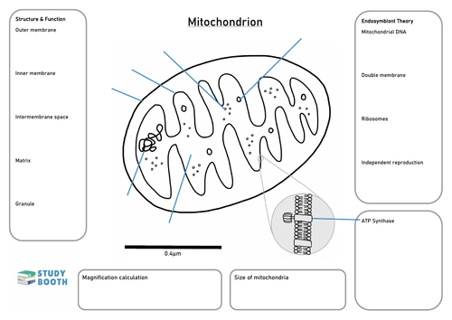
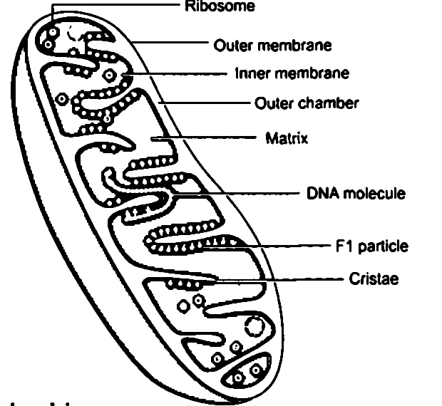
Parts of a Mitochondria Diagram | An Overview Diagram of a mitochondria Each mitochondrion has its DNA and ribosomes. The ribosomes use the genetic information contained by the mitochondrial DNA to produce protein or enzymes. An enzyme... Mitochondrion A mitochondrion (/ ˌ m aɪ t ə ˈ k ɒ n d r i ə n /; PL mitochondria) is an organelle found in the cells of most eukaryotes, such as animals, plants and fungi.Mitochondria have a double membrane structure and use aerobic respiration to generate adenosine triphosphate (ATP), which is used throughout the cell as a source of chemical energy. They were discovered by Albert von Kölliker in ... A Labelled Diagram Of Mitochondria with Detailed Explanation Diagram Of Mitochondria The diagram below shows the structure and functions of the mitochondria. Structure and Functions Of Mitochondria Matrix It is a viscous or gel-like fluid containing a mixture of enzymes, ribosomes, inorganic ions, mitochondrial DNA, nucleotide cofactors, and organic molecules. Diagram of Mitochondria | Labelled Diagram of Mitochondria Mitochondria have a double-membrane structure and also contain their own DNA. Therefore the inner membrane of the mitochondria is folded into structures called cristae, which increase the surface area available for the production of ATP. Labelled Diagram of a Mitochondrion. The diagram of a mitochondrion shows the internal structure of the ...
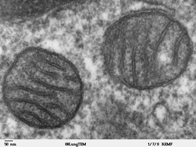
Animal cells - Cell structure - Edexcel - GCSE Biology (Single Science ... Mitochondria (singular: mitochondrion) are visible with a light microscope but can't be seen in detail. Ribosomes are only visible with an electron microscope. Preparing cheek cell slides to view ... Mitochondria - Structure - Function - TeachMePhysiology Mitochondria (singular: mitochondrion) are double membrane-bound cell organelles with a typical size of 0.75-3 μm². They are found in most mammalian cells, with notable exceptions including mature erythrocytes. 5.7.2 Structure of the Mitochondrion Structure of the Mitochondrion Mitochondria are rod-shaped organelles 0.5 - 1.0 µm in diameter They are the site of aerobic respiration in eukaryotic cells The function of mitochondria is to synthesize ATP Synthesis of ATP in the mitochondria occurs during the last stage of respiration called oxidative phosphorylation Mitochondria- Definition, Structure, Functions and Diagram Figure: Diagram of Mitochondria Structure of Mitochondria Mitochondria are mobile, plastic organelles that have a double-membrane structure. It ranges from 0.5 to 1.0 micrometer in diameter. It has four distinct domains: the outer membrane, the inner membrane, the intermembrane space, and the matrix.
Mitochondria Labeling Diagram | Quizlet
Mitochondria - Definition, Function & Structure | Biology Dictionary Structure of Mitochondria. Mitochondria have two membranes, an outer membrane and an inner membrane. These membranes are made of phospholipid layers, just like the cell's outer membrane. The outer membrane covers the surface of the mitochondrion, while the inner membrane is located within and has many folds called cristae. The folds increase ...

Draw a neat and labelled diagram showing ultra structure of ...
Animal cells - Cell structure - AQA - GCSE Combined Science Revision ... Below the basic structure is shown in the same animal cell, on the left viewed with the light microscope, and on the right with the transmission electron microscope. Mitochondria are visible...

Organ Sela Organelles Anatomi Biologis Vektor Diagram ...
Mitochondria Labeling Diagram Definition The inner membrane is also loaded with proteins involved in electron transport and ATP synthesis. This membrane surrounds the mitochondrial matrix, where the citric acid cycle produces the electrons that travel from one protein complex to the next in the inner membrane. Location Term Outer Membrane Definition

Structure of Mitochondria in 2023 | Biology diagrams, Cell ...
Plant cells - Cell structure - AQA - GCSE Combined Science Revision ... A tiny organelle where protein synthesis occurs. Plant cells also have additional structures: Function. Chloroplast. Organelles that contains the green pigment, chlorophyll, which absorbs light ...
Draw a neat and labelled diagram of mitochondria. - Sarthaks ...
Plant vs animal cells review A plant cell contains a large, singular vacuole that is used for storage and maintaining the shape of the cell. In contrast, animal cells have many, smaller vacuoles. Plant cells have a cell wall, as well as a cell membrane. In plants, the cell wall surrounds the cell membrane. This gives the plant cell its unique rectangular shape.

Structure & Function of Mitochondria (12.2.1) | CIE A Level ...
Mitochondria and chloroplasts The mitochondria are suspended in the jelly-like cytosol of the cell. They are oval-shaped and have two membranes: an outer one, surrounding the whole organelle, and an inner one, with many inward protrusions called cristae that increase surface area.
A Labelled Diagram Of Mitochondria with Detailed Explanation

How to draw mitochondria diagram easily | Mitochondria Diagram | YoKIdz | diagram of mitochondria

Draw a labelled diagram of a mitochondrion Why it is called a ...
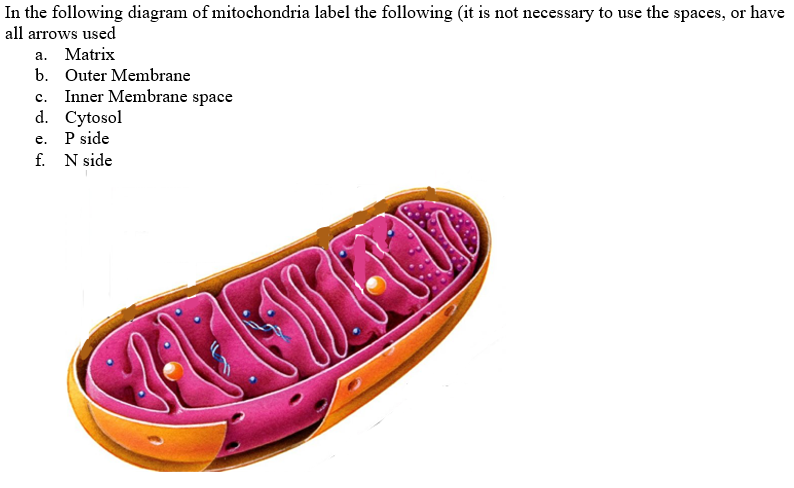
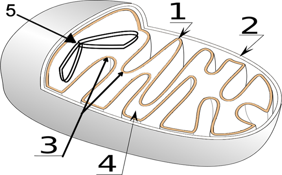
Solved In the following diagram of mitochondria label the ...
Mitochondria: Definition, Structure, Functions
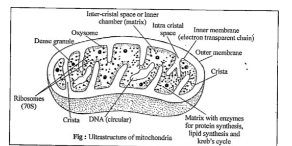
With the help of diagram explain the ultrastructure and ...
Mitochondria - Definition, Function & Structure | Biology ...

More about mitochondria — Science Learning Hub
Diagram of Mitochondria and Function of Mitochondria Parts ...

Structure Of Mitochondria || How To Draw And Label Mitochondria || Biology

Mitochondria: Form, function, and disease
Mitochondrion - Wikipedia
How Does the Mitochondria Produce Energy for the Cell
0614 Mitochondrion biology Medical Images For PowerPoint ...
Mitochondria worksheet - A Level Biology | Teaching Resources
Mitochondria and chloroplasts (article) | Khan Academy

Premium Vector | Diagram of the structure of mitochondria ...
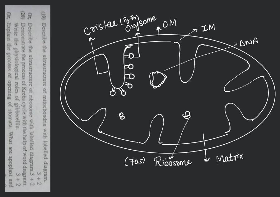
19) Describe the ultrastructure of mitochondria with labelled ...

Labeling the Mitochondria Diagram | Quizlet

how to draw diagram of mitochondria step by step | how to draw labelled diagram of mitochondria

How to draw Mitochondria | Well labelled diagram of Mitochondria | By Chetan Pachauri

Mitochondria | Definition, Examples, Diagrams

How to draw mitochondria | Diagram | Easy and well labelled diagram |NCERT | Power house of the cell

558 Chloroplast Mitochondria Images, Stock Photos & Vectors ...

Describe the structure of mitochondria with the help of a ...
Draw a neat labelled diagram of mitochondria. - Sarthaks ...

DRAW IT NEAT: How to draw Mitochondria

What Is Mitochondria (Structure, Diagram & Function)
Draw a neat labelled diagram of mitochondria. - Sarthaks ...
Solved] label two parts of the mitochondria. Label where each ...
The Fundamental Unit Of Life Class 9 Science Chapter 5 — CBSE ...
Draw and label the structure of Mitochondira.

draw a neat labelled diagram of mitochondria.help me out how ...
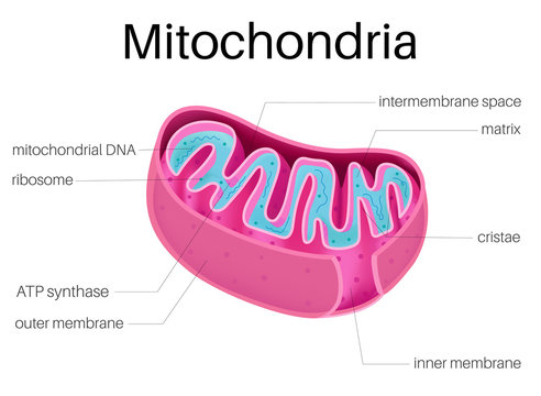
Illustration diagram of Mitochondria Stock Vector | Adobe Stock
Post a Comment for "40 labelled diagram of mitochondria"