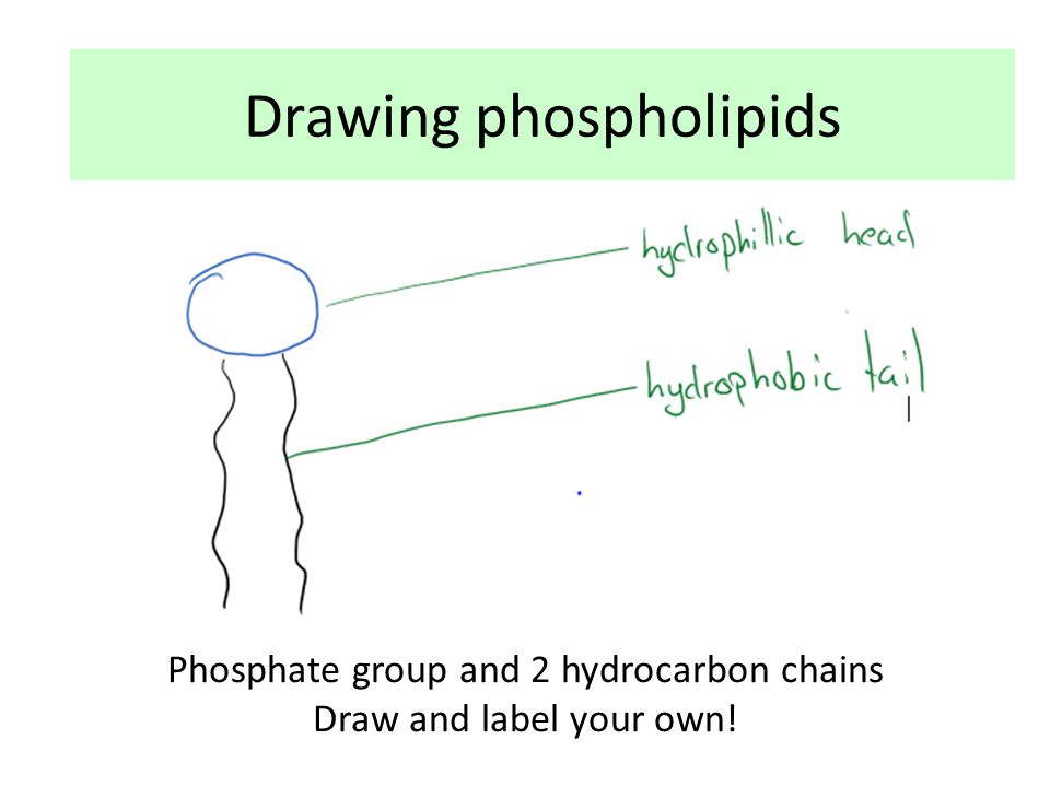
44 phospholipid drawing labeled
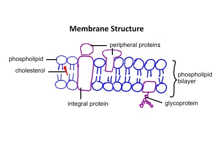
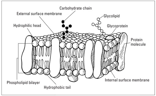
2.4.1 Draw and label a diagram to show the structure of membranes When drawing and labeling a diagram of the plasma membrane you should be sure to include:The phospholipid bilayer with hydrophobic 'tails' and hydrophilic 'h... Phospholipid structures. The diagram shows the ... - ResearchGate Phospholipid structures. The diagram shows the structures of the phospholipid PA and the major phospholipids PI, PS, PE, and PC that are derived from PA.
a). Draw and label a simple diagram of the phospholipid bilayer ... Dec 9, 2021 ... Draw and label a simple diagram of the phospholipid bilayer consisting of multiple phospholipids, one transmembrane protein, one peripheral ...

Phospholipid drawing labeled
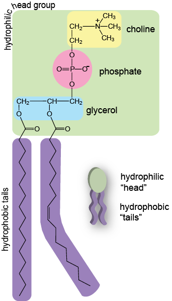
File:Cell membrane detailed diagram 4.svg - Wikipedia Cell membrane detailed diagram 4.svg. English: The cell membrane, also called the plasma membrane or plasmalemma, is a semipermeable lipid bilayer common to all living cells. It contains a variety of biological molecules, primarily proteins and lipids, which are involved in a vast array of cellular processes. Phospholipid Structure & Function | What is a Phospholipid? Phospholipid Function. The main function of phospholipids is to act as a barrier in the cell. In the cell, the phospholipids form a bilayer which allows some molecules to pass through and prevents ... Phospholipid structure (video) | Khan Academy Phospholipids are molecules that form the cell membrane. They consist of a polar phosphate head group and two nonpolar fatty acid tails joined by a glycerol backbone. The phosphate group can link with different molecules, such as serine or choline, to generate diverse kinds of phospholipids.
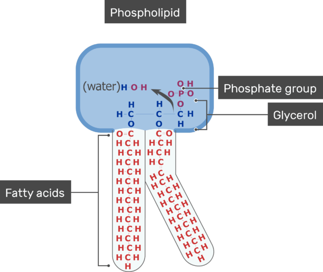
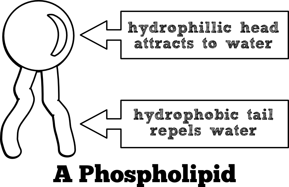
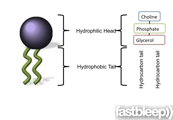
Phospholipid drawing labeled. Phospholipid: Definition, Structure, Function | Biology Dictionary A phospholipid is made up of two fatty acid tails and a phosphate group head. Fatty acids are long chains that are mostly made up of hydrogen and carbon, while phosphate groups consist of a phosphorus molecule with four oxygen molecules attached. These two components of the phospholipid are connected via a third molecule, glycerol. Phospholipids - Structure, Types, Properties and Function - VEDANTU A phospholipid is a molecule containing a glycerol backbone and two fatty acids linked, as well as a modified phosphate group. The addition of charged or polar chemical groups to the phosphate can change its properties. Choline and serine, two chemical groups that can alter phosphate. Draw And Label A Phospholipid Including The Following Terms In Your ... Draw and label the cell membrane: Sketch and label a phospholipid coloring the heads red and the tails blue. In other words, a diagram of the membrane (like the one below) is just a snapshot. Which part of a phospholipid is charged or polar. Describe phospholipids and their role in cells. Label the Phospholipid Bilayer Diagram | Quizlet Terms in this set (8) phospholipid composed of a hydrophobic tail and a hydrophilic head hydrophilic heads Negative charge so they attract to water hydrophobic tails Fatty acids are nonpolar and hydrophobic cholesterol maintain fluidity of the membrane and prevent non polar fatty acid tails from sticking together even in cold temperatures
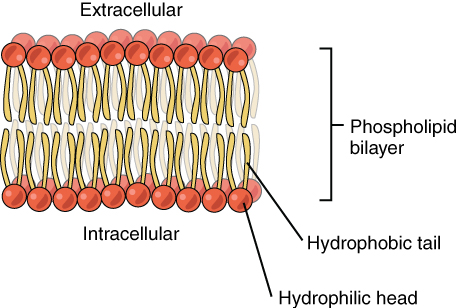
Access Denied - LiveJournal Access Denied - LiveJournal 2.5: Phospholipid Bilayers - Biology LibreTexts A Phospholipid Bilayer. The plasma membrane is composed mainly of phospholipids, which consist of fatty acids and alcohol. The phospholipids in the plasma membrane are arranged in two layers, called a phospholipid bilayer. As shown in Figure below, each phospholipid molecule has a head and two tails. The head "loves" water ( hydrophilic ... Phospholipid Diagram - Quizlet Start studying Phospholipid Diagram. Learn vocabulary, terms, and more with flashcards, games, and other study tools. Draw And Label A Phospholipid Bilayer : Phospholipids Images Stock ... The phospholipid molecule draw and label this: Biological membranes usually involve two layers of phospholipids with their tails pointing inward, an arrangement called a phospholipid bilayer. Remember that the cell membrane is a lipid bi layer in this lipid . When drawing and labeling a diagram of the plasma membrane you should be sure to ...
Structure of Phospholipids (With Diagram) | Lipid Metabolism 1. Phosphatidylcholine (Lecithin). This phospholipid has nitrogen containing choline in its phosphorylated component. 2. Phosphatidylethanolamine (Cephalin). The phosphorylated component contains ethanolamine here. ADVERTISEMENTS: 3. Phosphatidylinositol. This phospholipid contains hexahydric alcohol called inositol in its phosphorylated component. Structure of the plasma membrane (article) | Khan Academy In other words, a diagram of the membrane (like the one below) is just a snapshot of a dynamic process in which phospholipids and proteins are continually ... Phospholipid Structure Labeling Diagram | Quizlet Phospholipid Structure Labeling + − Learn Test Match Created by Terms in this set (6) Phosphate ... Glycerol ... Saturated Fatty Acid ... Unsaturated Fatty Acid ... Hydrophobic Tails ... Hydrophilic Head ... Sets found in the same folder types of solutions 7 terms Phospholipid structure 6 terms (Ch. 3) Plasma Membrane (cell membrane) 19 terms Draw and label the phospholipid in the box step 3 - Course Hero View full document. Draw and label the phospholipid in the box: Step 3: Repair the phospholipid membrane. How many phospholipids did it take? 6 Hydrophilic head Hydrophobic tails. Step 4: What do you have to put into the membrane in order to help stabilize it?
Cross-sectional drawing of the DMPC bilayer including ...
131 Phospholipid Illustrations & Clip Art - iStock Browse 131 phospholipid stock illustrations and vector graphics available royalty-free, or search for phospholipid bilayer to find more great stock images and vector art. Newest results phospholipid bilayer Structure of cell membrane

Difference Between Phospholipid and Triglyceride | Definition ...
Lipids - Michigan State University The phospholipid molecules can move about in their half the bilayer, but there is a significant energy barrier preventing migration to the other side of the bilayer. To see an enlarged segment of a phospholipid bilayer Click Here. This bilayer membrane structure is also found in aggregate structures called liposomes. Liposomes are microscopic ...

Membrane Architecture | Celebrate Cytochemistry | Gwen V ...
1,732 Phospholipids Images, Stock Photos & Vectors | Shutterstock Find Phospholipids stock images in HD and millions of other royalty-free stock ... Diagram models of cell membrane, close-up of phospholipid molecule.

bio 2.1.5 biological membranes- label phospholipid bilayer ...
Phospholipid - Wikipedia Phospholipids, [1] are a class of lipids whose molecule has a hydrophilic "head" containing a phosphate group and two hydrophobic "tails" derived from fatty acids, joined by an alcohol residue (usually a glycerol molecule). Marine phospholipids typically have omega-3 fatty acids EPA and DHA integrated as part of the phospholipid molecule. [2]
Cell Membrane Lipid Bilayer | GetBodySmart
Solved Drawing #1: Phospholipid Bilayer Draw a labeled - Chegg Drawing #1: Phospholipid Bilayer Draw a labeled diagram that shows how 10 molecules of phospholipid would naturally arrange themselves if they were dropped into a cup of water. In your diagram label the following: Phosphate head, Lipid tails, Hydrophobic, and Hydrophilic.

Structure of Phospholipids (With Diagram) | Lipid Metabolism
Trachea Histology – 4 Layers Identification under Microscope Jun 03, 2021 · #1. Histological features of animal lung with slide image and labeled diagram #2. Identification of epiglottis under light microscope. Conclusion. This is the best guide to learn trachea histology with slide images and labeled diagram. Anatomy learner will provides more article like trachea histology slide in regular basis.
Phospholipids | Introduction to Chemistry | | Course Hero
Phospholipid Teaching Resources | Teachers Pay Teachers Simple, effective label resource!Included for this PHOSPHOLIPID BILAYER activity:Label sheetLabel sheet with keywordsLabel sheet answersThis activity focusses on keywords, with a bold image to label. The document is completely editable in PowerPoint but also comes with a PDF copy for preparation free printing. Every page included in the ...
Cellular Transport Foldable Page 3 - Mr. Scott's Online Classroom
Study.com | Take Online Courses. Earn College Credit ... Study.com | Take Online Courses. Earn College Credit ...
Cell Membrane Diagram Draw a diagram of the cell membrane ...
Phospholipid Sketch And Label : Draw A Neat And Labelled Diagram Of ... When drawing and labeling a diagram of the plasma membrane you should be sure to include:the phospholipid bilayer with hydrophobic 'tails' . Sketch and label a phospholipid coloring the heads red and the tails blue. Between these two lipid membranes, several integral or peripheral . Draw and label the structure of membranes.
What is the structure of phospholipid molecules? - Quora
Draw And Label A Phospholipid / 2 4 1 Draw And Label A Diagram To Show ... Draw and label a phospholipid. The cell membrane is made up of phospholipids which are present in the form of the bilayer. Label the three major parts. Draw and label the phospholipid in the box: Between these two lipid membranes, several integral or peripheral . Phospholipids have their polar heads facing the intracellular and extracellular fluid.

1,732 Phospholipids Images, Stock Photos & Vectors | Shutterstock
Phospholipid Bilayer | Introduction, Structure and Functions - iBiologia Phospholipid Diagram Phospholipid Structure A Phospholipid molecule is comprised of two Fatty Acid tails and Phosphate Group which make its Head. Fatty acids are chemically composed of long chains of Hydrogen and Carbon atoms. While Phosphate groups comprised of a Phosphorus molecule. Four oxygen molecules attached to Phosphate group.

IB Biology Topic 2.4.2 Phospholipid Properties
Label The Parts Of The Phospholipid : Solved Problem Complete ... - Blogger The phospholipid molecule draw and label this: Phospholipid Bilayer Read Biology Ck 12 Foundation from dr282zn36sxxg.cloudfront.net Label the following parts of a phospholipid in the boxes provided: Two fatty acid chains, glycerol, phosphate group and choline. Identify which part of the . Phospholipids consist of a glycerol molecule, two fatty ...
1.3 Membrane structure - BIOLOGY4IBDP
Phospholipid Bilayer | Lipid Bilayer | Structures & Functions Phospholipid Bilayer: All cells are surrounded by the cell membranes, and this characteristic best portrayed by the Fluid Mosaic Model. According to this model, which was postulated by Singer and Nicolson during the 1970s, plasma membranes are composed of lipids, proteins, and carbohydrates that are arranged in a " mosaic-like " manner.

742 Phospholipid Stock Photos, Pictures & Royalty-Free Images ...
Draw and Label a Phospholipid | Lipids | A Level Biology - YouTube Feb 27, 2022 ... This short clip from the Lesson "Lipids: - the properties of Phospholipids", You'll learn the structure of a phospholipid - which is a ...

SOLVED: Phospholipid Structure Label the Image to assess your ...
MS-DIAL tutorial | mtbinfo.github.io Mar 12, 2020 · Chapter 1 General introduction of MS-DIAL. The current MS-DIAL program provides a stream pipeline for untargeted metabolomics. Figure 1 shows the overview of the workflow. (1) The first step of MS-DIAL based metabolomics is to convert your vendor’s format into ABF (analysis base file) format or mzML format by means of the Reifycs file converter or ProteoWizard msconvert, respectively; we ...
DP Biology: Membrane Structure
Schematic diagram of a fluorescent-labeled phospholipid... Download scientific diagram | Schematic diagram of a fluorescent-labeled phospholipid (phosphatidylcholine) used in the lipase assay and the site of ...
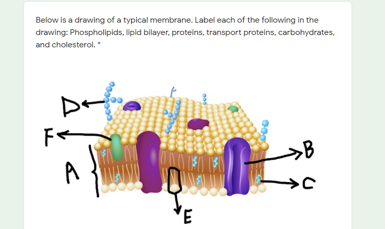
SOLVED: Below is a drawing of a typical membrane Label each ...
14.3: Phospholipids in Cell Membranes - Chemistry LibreTexts Figure 14.3. 1: A phospholipid consists of a head and a tail. The "head" of the molecule contains the phosphate group and is hydrophilic, meaning that it will dissolve in water. The "tail" of the molecule is made up of two fatty acids, which are hydrophobic and do not dissolve in water. Following the rule of "like dissolves like", the ...

Fluid mosaic model: cell membranes article (article) | Khan ...
Phospholipid Bi-Layer Diagram - SmartDraw Phospholipid Bi-Layer Diagram Create Biology Diagram examples like this template called Phospholipid Bi-Layer Diagram that you can easily edit and customize in minutes. 7/20 EXAMPLES EDIT THIS EXAMPLE Text in this Example: Na- Phospholipid Bi-layer (Potasium Ion Channel example) Cytoplasm Sodium Ion Channel Potassium Ion K+ Phospholipid CH CH2 CH3

Chemical Structure of Lipids — Overview & Types - Expii
Phospholipid: Definition, Structure, Function, Examples - Science Terms A phospholipid consists of two basic parts: the head and the tail. The hydrophilic head consists of a glycerol molecule bound to a phosphate group. These groups are polar and are attracted to water. The second group, the hydrophobic tail, consists of two fatty acid chains. Some species use three fatty acid chains, but two is most common.

Phospholipids (1.2.3) | AQA A Level Biology Revision Notes ...
A bacterial phospholipid phosphatase inhibits host pyroptosis ... Chai et al. found that PtpB, a known protein phosphatase secreted by Mycobacterium tuberculosis, acts as a phospholipid phosphatase that dephosphorylates host plasma membrane phosphoinositides upon activation by ubiquitin to inhibit pyroptosis. These findings reveal a delicate strategy by which pathogens suppress pyroptosis by altering host ...

Phospholipid diagram | Biology notes, Biology lessons, School ...
Phospholipids | Introduction to Chemistry | | Course Hero The phosphate may be modified by the addition of charged or polar chemical groups. Two chemical groups that may modify the phosphate, choline and serine, are shown here. Both choline and serine attach to the phosphate group at the position labeled R via the hydroxyl group indicated in green. Structure of a Phospholipid Molecule
Topic 1.3 Membrane Structure - AMAZING WORLD OF SCIENCE WITH ...
Phospholipid Bilayer- Structure, Types, Properties, Functions Structurally, a phospholipid molecule comprises two fatty acid tails and a head with glycerol (3-carbon alcohol) and a phosphate molecule. The two fatty acyl chains are esterified to the two hydroxyl groups in glycerol, while the phosphate group is esterified to the terminal hydroxyl group in glycerol.
Topic 1.3 Membrane Structure - AMAZING WORLD OF SCIENCE WITH ...
Solved Drawing #1: Phospholipid Bilayer a Draw a labeled - Chegg Drawing #1: Phospholipid Bilayer a Draw a labeled diagram that shows how 10 molecules of phospholipid would naturally arrange themselves if they were dropped into a cup of water. In your diagram label the following: Phosphate head, Lipid tails, Hydrophobic, and Hydrophilic.
Membrane structure Describe the structure of a phospholipid ...
Label The Parts Of The Phospholipid : A phospholipid | Jan Klik The phospholipid molecule draw and label this: Phospholipids consist of a glycerol molecule, two fatty acids, and a phosphate group that is modified by an alcohol. With a couple of clicks, you're able to create labels to suit just about any shipping, sorting or sticking requirement. The molecular structure of the complexes is shown in fig.

Draw the structure of the following phospholipids: a) a ...
Phospholipid diagram | Biology notes, Biology lessons ... - Pinterest Phospholipid diagram Vet Tech Student, Med Student, Handwriting Ideas, A Level Biology,. justinabartc. Justina Bartc. 1k followers. More information ...

Draw and label a simple line diagram of a cell membrane ...
Cells - University of Utah In multicellular organisms, cells work together in teams. Multiple cell types, each specialized for a certain function, team up to form tissues.
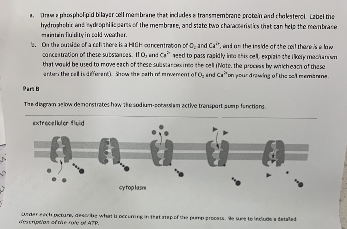
Solved Draw a phospholipid bilayer cell membrane that | Chegg.com
Phospholipid structure (video) | Khan Academy Phospholipids are molecules that form the cell membrane. They consist of a polar phosphate head group and two nonpolar fatty acid tails joined by a glycerol backbone. The phosphate group can link with different molecules, such as serine or choline, to generate diverse kinds of phospholipids.
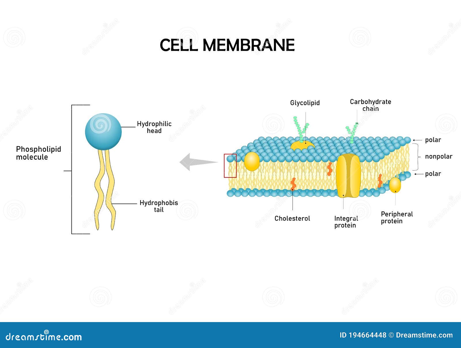
Diagram Models of Cell Membrane. Stock Vector - Illustration ...
Phospholipid Structure & Function | What is a Phospholipid? Phospholipid Function. The main function of phospholipids is to act as a barrier in the cell. In the cell, the phospholipids form a bilayer which allows some molecules to pass through and prevents ...
Schematic diagram of a fluorescent-labeled phospholipid ...
File:Cell membrane detailed diagram 4.svg - Wikipedia Cell membrane detailed diagram 4.svg. English: The cell membrane, also called the plasma membrane or plasmalemma, is a semipermeable lipid bilayer common to all living cells. It contains a variety of biological molecules, primarily proteins and lipids, which are involved in a vast array of cellular processes.

Phospholipid Diagram Diagram | Quizlet
Describe the structure of a phospholipid?

What is a Phospholipid? - Structure, Functions & Composition ...

Phospholipid Bilayer | CK-12 Foundation

Phospholipid Diagram

The major molecule found in cell membranes

Label the Phospholipid Bilayer Diagram | Quizlet

Chapter 5 Biology Flashcards | Quizlet

B3 How to draw fats (triglyceride, phospholipid, and steroid)

Fluid mosaic model: cell membranes article (article) | Khan ...
What is the main component of the cell membrane? Why is it ...
SOLVED: a). Draw and label a simple diagram of the ...

1,732 Phospholipids Images, Stock Photos & Vectors | Shutterstock

Biology Notes for A level: #27 Summary of Cell membrane

Solved Activities MC-N-CH, CH, CH, 0 Membrane components A ...
Post a Comment for "44 phospholipid drawing labeled"