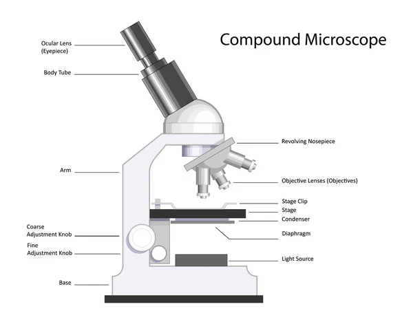
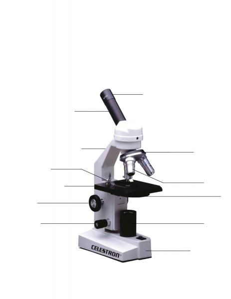
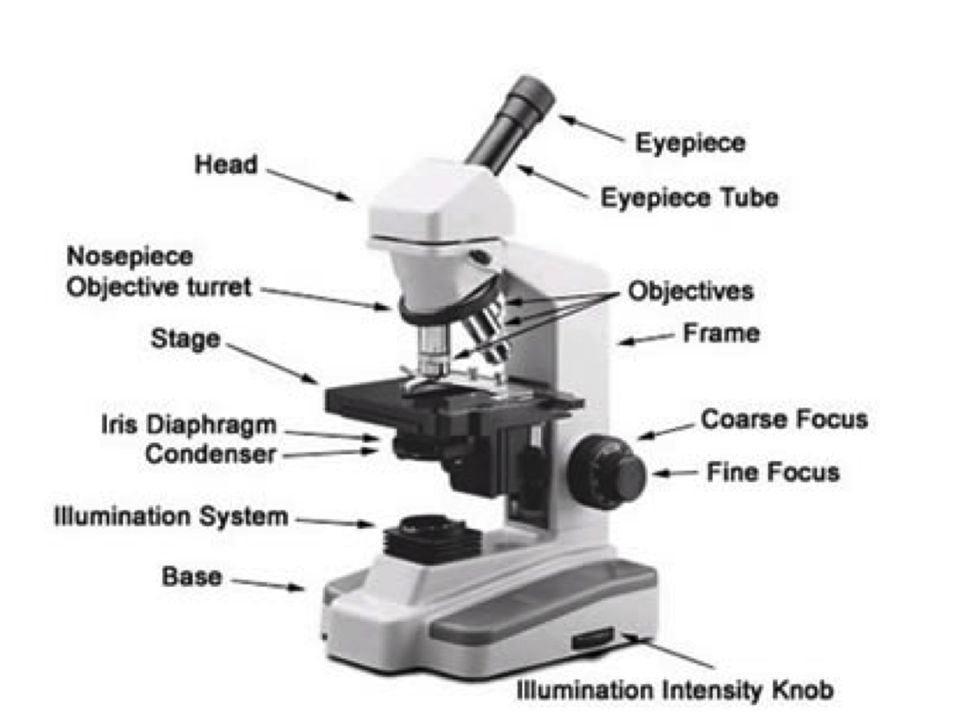
44 microscope picture with labels
The principle of STochastic Optical Reconstruction Microscopy Activator-free labels consist of imaging dye only, enabling simple labeling and sample preparation techniques such as conventional indirect immunofluorescence using conventional dye-conjugated antibodies. Product Brochure Download 11.11MB Super-Resolution Microscopes N-SIM S N-SIM E N-STORM STEDYCON Mr. Jones's Science Class Matter: Atoms and Properties - Open Response Question 3. Force and Motion - Open Response Question 3. Forms of Energy - Open Response Question 1. Forms of Energy - Open Response Question 2. Earth's Structure & Natural Processes - Open Response Question 1.
A Single-cell Morphological Dataset of Leukocytes from AML ... - Wiki The Munich AML Morphology Dataset contains 18,365 expert-labeled single-cell images taken from peripheral blood smears of 100 patients diagnosed with Acute Myeloid Leukemia at Munich University Hospital between 2014 and 2017, as well as 100 patients without signs of hematological malignancy.
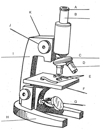
Microscope picture with labels
Metaphase - Genome.gov Definition. Metaphase is a stage during the process of cell division (mitosis or meiosis). Normally, individual chromosomes are spread out in the cell nucleus. During metaphase, the nucleus dissolves and the cell's chromosomes condense and move together, aligning in the center of the dividing cell. At this stage, the chromosomes are ... principle of light microscope Home / カテゴリーなし / principle of light microscope. principle of light microscope. 2022-09-11T06:47:19+09:00 2022-09-11 | 2022-09-11 | Spin-selective tunneling from nanowires of the candidate topological ... Topological insulators exhibit so-called spin-momentum locking, which has been put to use in spintronics applications. Aishwarya et al. exploited these properties to achieve spin-polarized tunneling through a scanning tunneling microscope (STM) tip. The researchers mounted an SmB 6 nanowire onto a trimmed tungsten tip.
Microscope picture with labels. 5 White Blood Cells Types and Their Functions - New Health Advisor Agranulocytes are free of visible grains under the microscope and include lymphocytes and monocytes. Together, they coordinate with one another to fight off things like cancer, cellular damage, and infectious diseases. Below, detailed information about each type will be discussed. 1. Neutrophils LA shooting in May seems to match Cheyenne Floyd shooting details Another witness told a reporter she heard six shots. A photo of the bullet-ridden car seems to show at least eight bullet holes on the passenger side. You will notice that all of the apparent... DP Biology: Calculating Magnification and Size Activity 1 Calculating magnification of an image using it's scale bar The three images below (click the eye to reveal) show a worked example of how to calculate sizes of cells organelles from electron micrographs step by step. Follow these steps carefully then complete the calculations on the worksheet. Venn Diagram Templates | Editable Online or Download for Free 4 Set Venn diagram template. Venn Diagram of Cholesterol vs Blood Pressure. Venn Diagram Template on Student Behavior. Math Euler Diagram Template. Venn Diagram on Project Failure. 3 Circle Venn Diagram to Download or Modify Online. Drawing Venn diagram with Creately.
A new 3D tool for screening and measuring int | EurekAlert! A new publication from Opto-Electronic Advances; DOI 10.29026/oea.2023.220048 discuss a new 3D tool for screening and measuring intracellular lipid droplets using flow tomography. Refractive Index Sensing Performance of Stepped-mTFBG Fabricated by ... 1. Introduction. Among the various of optical fiber sensing technologies, fiber Bragg grating (FBG) has been playing the most important role for its practical applications , .The reflection signal with special interference wavelength (λ Bragg) can be demodulated to realize the real-time measuring temperature and strain, etc. .Furthermore, the quasi-distributed system can simultaneously and ... Light Microscope (Theory) - Amrita Vishwa Vidyapeetham Microscope Microscope is an optical instrument that uses lens or combination of lens to produce magnified images that are too small to seen by unaided eye. Microscope provides the enlarged view that helps in examining and analyzing the image. Enzymatic cascade reaction in simple-coacervates - ScienceDirect Bright field and fluorescence images were obtained using a Zeiss microscope with an objective ×50. For the samples labeled with Alexa Fluor 488, the excitation wavelength was 500 nm, for the samples labeled with resorufin, the excitation wavelength was 600 nm. The treatment was performed with the software AxioVision. 3. Results and discussion3.1.
Human Biology Lab Online | Lab 4 Tissues and Skin When viewing the slides, please follow these steps: Start on the lowest magnification 40x Move to the next higher magnification 100x View the tissue at 400x (Make your sketch of the tissue at this mag.) Can you identify any of the cell structures Select the Labels "On" button Use the slider bars on the side and bottom to scan around the slide. King Charles: What kind of monarch should we expect Elizabeth's son to ... As Prince of Wales, Charles has worn many labels. So the world should already have a fair idea of what sort of king Charles III will be. And how, or if, the monarchy will change under him. Director Alice Diop on Her Venice Film Festival Winner 'Saint Omer' Saint Omer. , Director Alice Diop Puts Motherhood Under the Microscope. The director won the Silver Lion Grand Jury prize at the Venice Film Festival. by Nicolas Rapold. 09.12.22. Alice Diop wears ... Skin Layers: Structure, Function, Anatomy, and More - Verywell Health The skin is the body's largest organ. It is made of three layers, each of which has specific functions. The outermost epidermis is responsible for producing new skin cells, protecting the body from unwanted substances, and retaining moisture to keep the skin well hydrated. The middle dermis is responsible for supporting and strengthening the skin.
The Compound Light Microscope Label the following parts on ...
Fluorescence In Situ Hybridization (FISH) - Genome.gov Fluorescence in situ hybridization (abbreviated FISH) is a laboratory technique used to detect and locate a specific DNA sequence on a chromosome. In this technique, the full set of chromosomes from an individual is affixed to a glass slide and then exposed to a "probe"—a small piece of purified DNA tagged with a fluorescent dye.

Parts of a Microscope with Their Functions • Microbe Online
A diamond voltage imaging microscope | Nature Photonics Figure 3a shows a scanning electron microscopy image of a diamond sensor surface with an array of 700 nm-diameter pillars produced by reactive ion etching (Methods). As evidenced by Fig. 3b, these...

Microscope With Labels clip art | Microscope parts ...
Quiz: Label The Parts Of The Eye - ProProfs Quiz Quiz: Label The Parts Of The Eye. Do you know the anatomy of the human eye very well? Can you label the parts of the eye in the quiz below? Give it a try and evaluate yourself. The eye has many important parts, each with different functions, including the cornea, pupil, sclera, and many more. Can you tell where these parts are located and what ...

Labeling a Microscope Free Worksheet Pack
AX R MP | Multiphoton Microscopes | Nikon Microscope Products | Nikon ... Use the AX R MP field of view and Nikon's world-class objective lenses to capture large specimens in one image. With resolutions up to 8K x 8K, these images can be captured without sacrificing a single detail. Redesigned optics maximize performance over a large area and deliver seamless image stitching at much higher speeds.

Parts of a microscope with functions and labeled diagram
Gram Staining Procedure | New Health Advisor 2. Label the Slides. Draw a circle under the slides using a marking pen designed for glassware. This will help to designate which area to prepare the smear in the following step. You can also label them with the organism's initials at the edge of each slide. Take care that the labels do not get in contact with the reagentsused forstaining. 3.

Biology label part of microscope
An Expert-Annotated Dataset of Bone Marrow Cytology in ... - Wiki Image acquisition was performed using a brightfield microscope with 40x magnification and oil immersion. Large datasets with a high quality of both data acquisition and annotation are key prerequisites to develop data-driven, computational methods in diagnostic medicine.
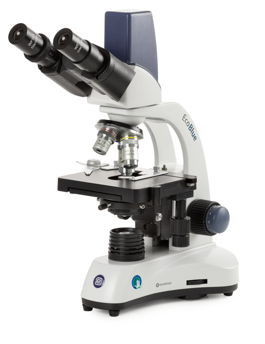
EcoBlue digital - Euromex
Light Microscope (Assignment) - Amrita Vishwa Vidyapeetham To illustrate the working of a microscope, follow the instructions in the simulator. Students Assignment . Are there any other objective used in light microscope other than 4X, 10X, 40Xand 100X? If YES, specify them? Consider a light microscope without iris diaphragm. How the images at 4 X, 10 X, 40 X and 100 X differ?

Jual Mikroskop Binokuler Microscope Binocular XSZ 107 BN XSZ ...
Liquid-interface assisted SERS could see earlier detection of Alzheimer ... (a) Schematic of the fabrication (b) Photograph of microfluidic SERS chip (c) Optical microscope image showing the SERS substrate. SEM images of (d) original metal film, (e) ripples generated by...
Transmitted light microscope B3 Professional series B3-220ASC ...
Diatom - Wikipedia Diatom (Neo-Latin diatoma) refers to any member of a large group comprising several genera of algae, specifically microalgae, found in the oceans, waterways and soils of the world.Living diatoms make up a significant portion of the Earth's biomass: they generate about 20 to 50 percent of the oxygen produced on the planet each year, take in over 6.7 billion metric tons of silicon each year from ...
4,814 Microscope labeled Images, Stock Photos & Vectors ...
Bacteriology Notes - Microbe Notes Bacteriology. Bacteriology is a branch or discipline of science that studies different characteristics of bacteria and their association with other organisms or disciplines. Over the years, the discipline of bacteriology has evolved from the microbiological tests performed only by physicians, the application of the germ theory of disease and ...

Label the light microscope | Teaching Resources
Event-driven acquisition for content-enriched microscopy In our implementation, the open software Micro-Manager 20 captures images from the microscope. As new frames are received, a neural network trained on labeled bio-image data to recognize specific...

National D-ELDB Digital Compound Binocular Microscope
Spin-selective tunneling from nanowires of the candidate topological ... Topological insulators exhibit so-called spin-momentum locking, which has been put to use in spintronics applications. Aishwarya et al. exploited these properties to achieve spin-polarized tunneling through a scanning tunneling microscope (STM) tip. The researchers mounted an SmB 6 nanowire onto a trimmed tungsten tip.
The Microscope
principle of light microscope Home / カテゴリーなし / principle of light microscope. principle of light microscope. 2022-09-11T06:47:19+09:00 2022-09-11 | 2022-09-11 |
Microscope | Other Quiz - Quizizz
Metaphase - Genome.gov Definition. Metaphase is a stage during the process of cell division (mitosis or meiosis). Normally, individual chromosomes are spread out in the cell nucleus. During metaphase, the nucleus dissolves and the cell's chromosomes condense and move together, aligning in the center of the dividing cell. At this stage, the chromosomes are ...
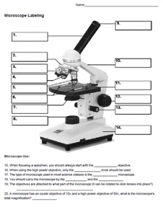
Microscope Labeling

The Microscope

Label the microscope — Science Learning Hub

Optical microscope, microscope, angle, simple, label png ...
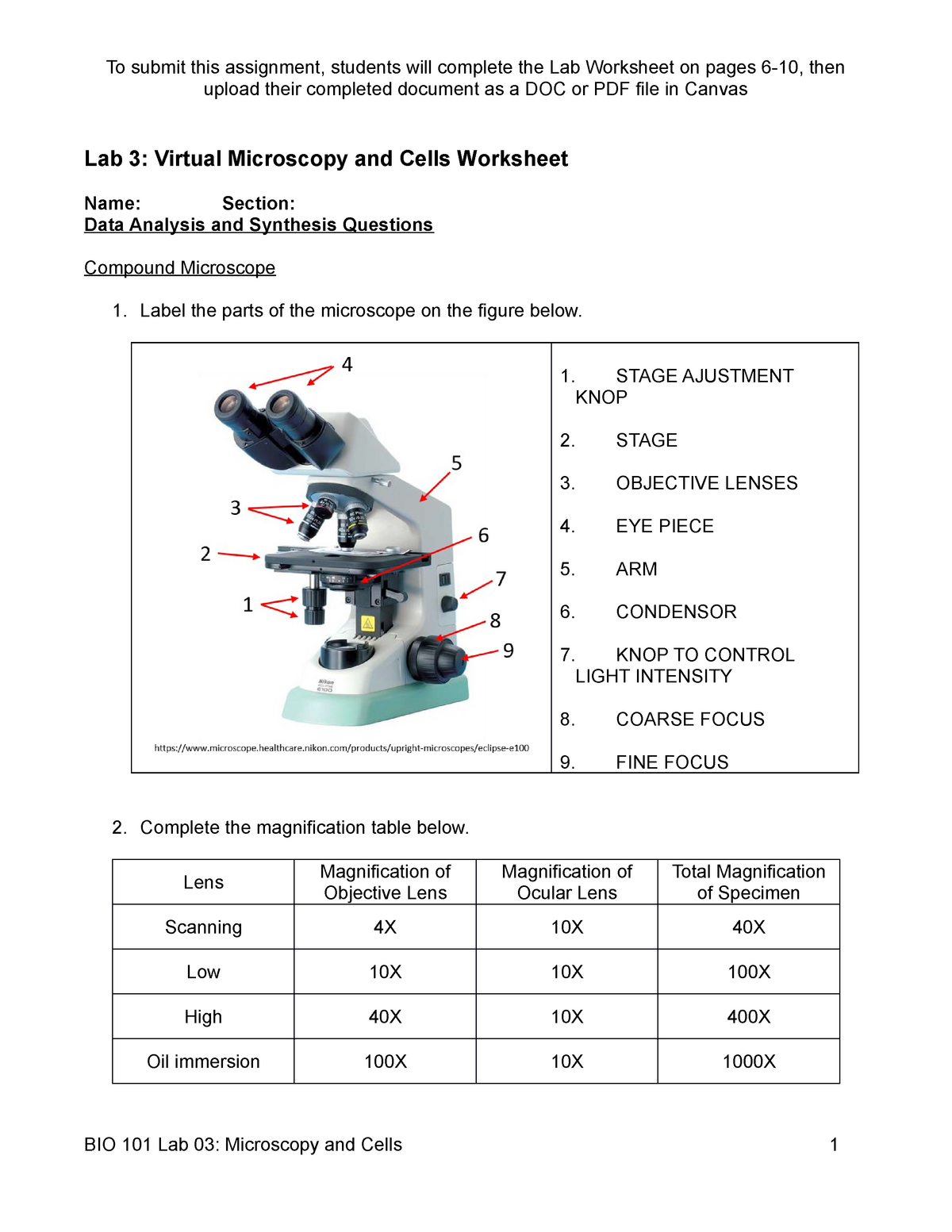
BIO 101 Lab 03, Microscopy and Cells,original - upload their ...

Microscope World Blog: Labeling the Parts of the Microscope
Microscope slide Vector Art Stock Images | Depositphotos
The Parts of a Microscope (Labeled) Printable Printable (6th ...

Cytology - BIO 1210: Human Anatomy and Physiology I ...

Microscope labeling
Label a microscope - Teaching resources

label the parts of microscope scope - Brainly.in

Microscope Labels Photos - Free & Royalty-Free Stock Photos ...
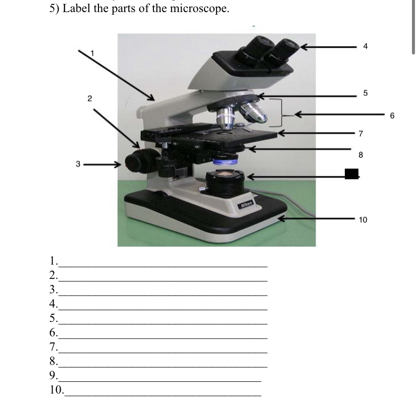
Answered: 5) Label the parts of the microscope. 1… | bartleby

Parts of a Light Microscope Activity | Labeling Task

Optical microscope - 037 Pro - Breukhoven - laboratory ...

Microscope Terms Glossary | Earth science lessons, Biology ...

MICROSCOPE Labeling - Part - 3

label microscope diagram | Charts | Microscope, Anatomy bones ...
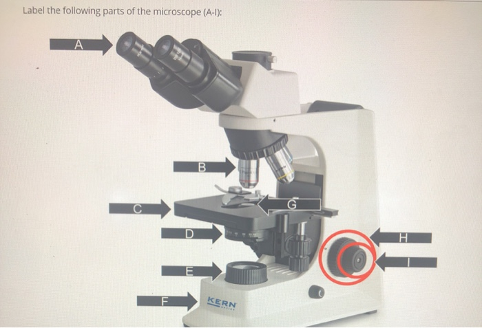
Solved Label the following parts of the microscope (A-1 ...
Lab - Microscope: MAH-Summer 2019-Anatomy and Physiology I

Microscope Diagram Labeled, Unlabeled and Blank | Parts of a ...

Microscope Labeling Practice Diagram | Quizlet

Parts of a microscope with functions and labeled diagram

Label the Microscope Diagram | Download Scientific Diagram

Below is a photo of a compound light microscope with labels ...
Label microscope - Teaching resources
Print Map Quiz: Labeling the Microscope ()

Activity 1).docx - Activity #1 THE MICROSCOPE I. A. Label the ...
Monday 10/19/15 AIM: how do the parts of the compound light ...
Post a Comment for "44 microscope picture with labels"