41 diagram of a microscope and label
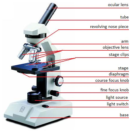
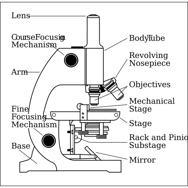
Microscope: Parts Of A Microscope With Functions And Labeled Diagram. Figure: A diagram of a microscope's components. The microscope has three basic components: the head, the base, and the arm. Head:Occasionally, the head is considered the body. It holds the optical components of the upper part of the microscope. Base:The microscope's base provides great support. Microscope Labeling Diagram | Quizlet Hold the slide in place on the stage. Nosepiece Holds the objective lenses and allows the lenses to rotate for viewing. Stage Supports the slide where the specimen is being viewed. Lamp Projects or reflects light upward through the diaphragm. Base Supports and stabilizes the microscope. Diaphragm
Label the Microscope Diagram | Download Scientific Diagram - ResearchGate Download scientific diagram | Label the Microscope Diagram from publication: Laboratory Exercises in Microbiology: Discovering the Unseen World through Hands-on Investigation | Microbiology ...

Diagram of a microscope and label
Family Tree Stock Photos, Pictures & Royalty-Free Images A set of genetics icons that include editable strokes or outlines using the EPS vector file. The icons include families, DNA, Genes, genetic testing concepts, cells, science and biology, microscope, geneticists, family tree, biologist using microscope, petri dish, human biology, human identity and other related icons. Microscope Types (with labeled diagrams) and Functions Simple microscope labeled diagram Simple microscope functions It is used in industrial applications like: Watchmakers to assemble watches Cloth industry to count the number of threads or fibers in a cloth Jewelers to examine the finer parts of jewelry Miniature artists to examine and build their work Also used to inspect finer details on products Compound Microscope Parts – Labeled Diagram and their … Major structural parts of a compound microscope. There are three major structural parts of a compound microscope. The head includes the upper part of the microscope, which houses the most critical optical components, and the eyepiece tube of the microscope.; The base acts as the foundation of microscopes and houses the illuminator.; The arm connects between the base …
Diagram of a microscope and label. en.wikipedia.org › wiki › Fluorescence_microscopeFluorescence microscope - Wikipedia "Fluorescence microscope" refers to any microscope that uses fluorescence to generate an image, whether it is a simple set up like an epifluorescence microscope or a more complicated design such as a confocal microscope, which uses optical sectioning to get better resolution of the fluorescence image. › 6-label-the-microscopeLabel the microscope — Science Learning Hub Jun 08, 2018 · All microscopes share features in common. In this interactive, you can label the different parts of a microscope. Use this with the Microscope parts activity to help students identify and label the main parts of a microscope and then describe their functions. Drag and drop the text labels onto the microscope diagram. If you want to redo an ... Compound Microscope - Diagram (Parts labelled), Principle and Uses See: Labeled Diagram showing differences between compound and simple microscope parts Structural Components. The three structural components include. 1. Head. This is the upper part of the microscope that houses the optical parts. 2. Arm . This part connects the head with the base and provides stability to the microscope. Microscope Parts, Function, & Labeled Diagram - slidingmotion Microscope parts labeled diagram gives us all the information about its parts and their position in the microscope. Microscope Parts Labeled Diagram The principle of the Microscope gives you an exact reason to use it. It works on the 3 principles. Magnification Resolving Power Numerical Aperture. Parts of Microscope Head Base Arm Eyepiece Lens
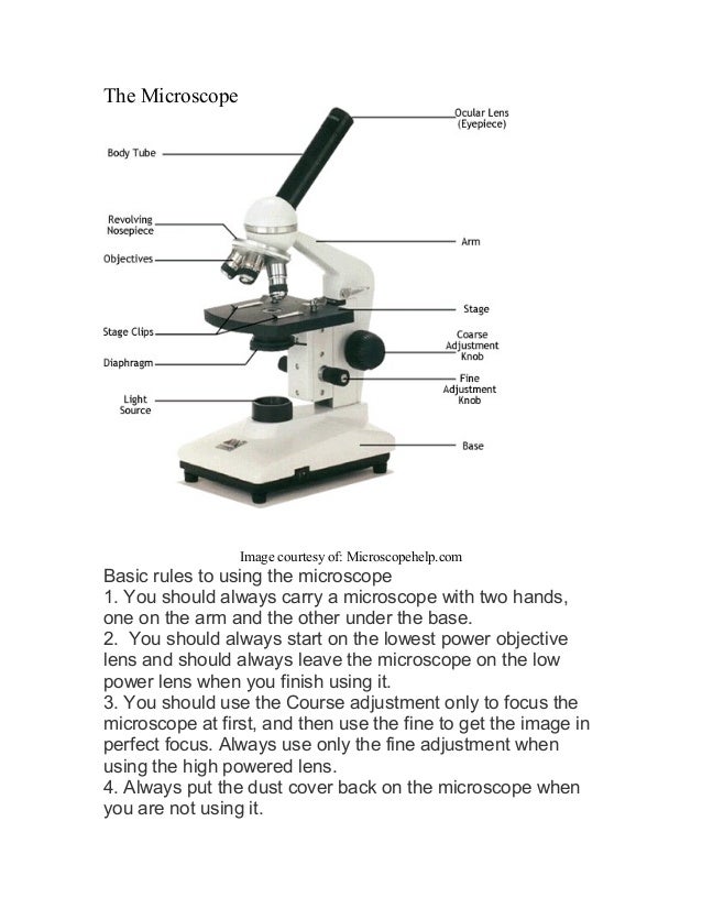
Microscope labeled diagram - slideshare.net Microscope labeled diagram 1. The Microscope Image courtesy of: Microscopehelp.com Basic rules to using the microscope 1. You should always carry a microscope with two hands, one on the arm and the other under the base. 2. You should always start on the lowest power objective lens and should always leave the microscope on the low power lens ... Sperm Under Microscope with Labeled Diagram - AnatomyLearner Sperm Under Microscope with Labeled Diagram 24/06/2022 17/06/2022 by anatomylearner While studying the histological features of the seminiferous tubules and epididymis, you will see sperm cells under the microscope. They are much smaller and lie in groups along the inner margin of the Sertoli cells. microbenotes.com › parts-of-a-microscopeParts of a microscope with functions and labeled diagram Apr 19, 2022 · Figure: Diagram of parts of a microscope. There are three structural parts of the microscope i.e. head, base, and arm. Head – This is also known as the body. It carries the optical parts in the upper part of the microscope. Base – It acts as microscopes support. It also carries microscopic illuminators. Labelled Diagram of Compound Microscope The below mentioned article provides a labelled diagram of compound microscope. Part # 1. The Stand: The stand is made up of a heavy foot which carries a curved inclinable limb or arm bearing the body tube. The foot is generally horse shoe-shaped structure (Fig. 2) which rests on table top or any other surface on which the microscope in kept.
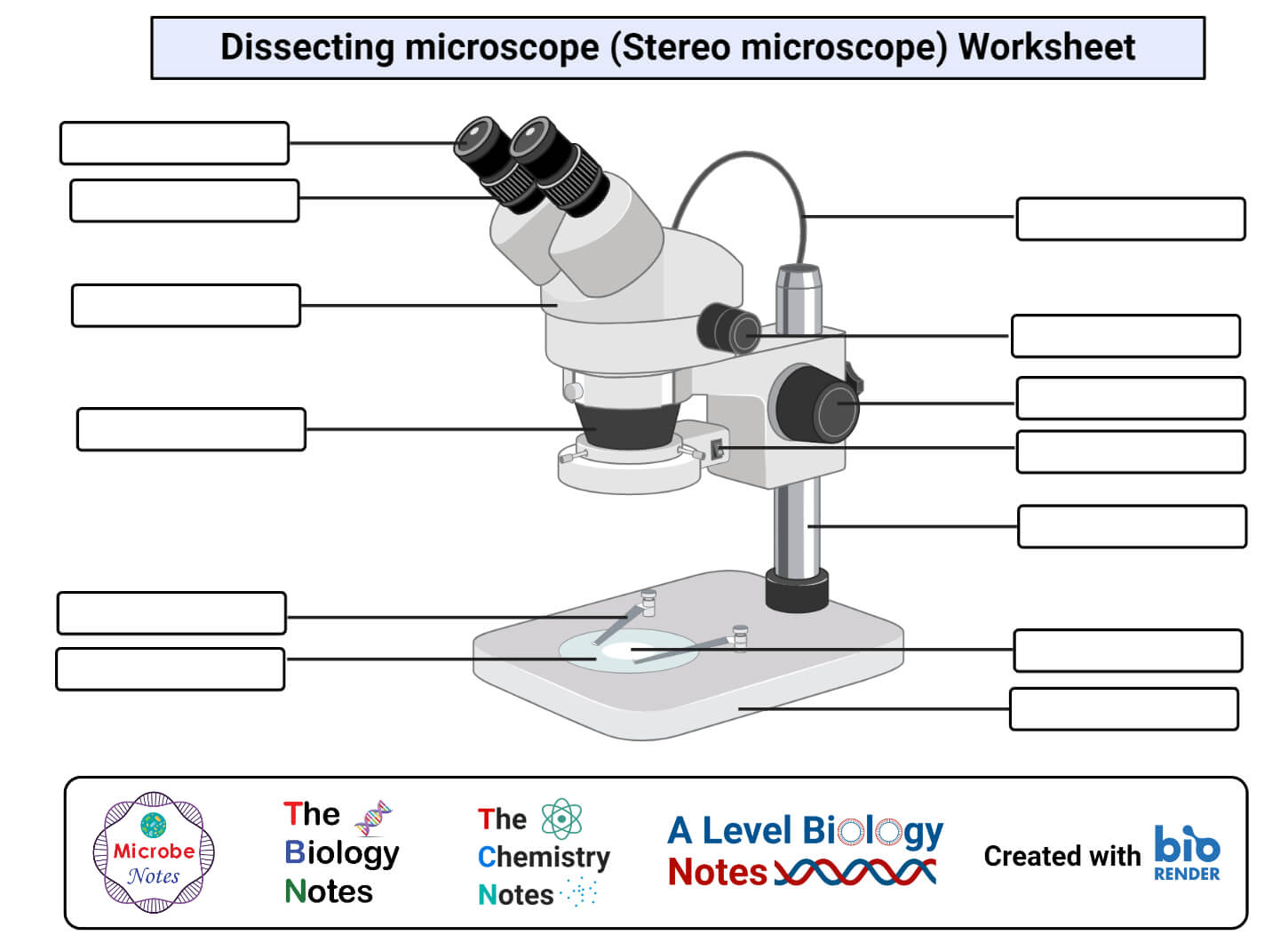
rsscience.com › stereo-microscopeParts of Stereo Microscope (Dissecting microscope) – labeled ... Labeled part diagram of a stereo microscope Major structural parts of a stereo microscope. There are three major structural parts of a stereo microscope. The viewing Head includes the upper part of the microscope, which houses the most critical optical components, including the eyepiece, objective lens, and light source of the microscope. Compound Microscope Parts - Labeled Diagram and their Functions There are three major structural parts of a compound microscope. The head includes the upper part of the microscope, which houses the most critical optical components, and the eyepiece tube of the microscope. The base acts as the foundation of microscopes and houses the illuminator. The arm connects between the base and the head parts. Microscope Parts and Functions First, the purpose of a microscope is to magnify a small object or to magnify the fine details of a larger object in order to examine minute specimens that cannot be seen by the naked eye. Here are the important compound microscope parts... Eyepiece: The lens the viewer looks through to see the specimen. Botulinum toxin - Wikipedia Botulinum toxin (BoNT), often shortened to Botox, is a neurotoxic protein produced by the bacterium Clostridium botulinum and related species. It prevents the release of the neurotransmitter acetylcholine from axon endings at the neuromuscular junction, thus causing flaccid paralysis. The toxin causes the disease botulism.The toxin is also used commercially for …
Compound Microscope Parts, Diagram Definition, Application ...
PDF Parts of a Microscope Printables - Homeschool Creations Label the parts of the microscope. You can use the word bank below to fill in the blanks or cut and paste the words at the bottom. Microscope Created by Jolanthe @ HomeschoolCreations.net. Parts of a eyepiece arm stageclips nosepiece focusing knobs illuminator stage objective lenses

Compound Microscope – Diagram (Parts labelled), Principle and ...
Parts of a microscope with functions and labeled diagram Apr 19, 2022 · Figure: Diagram of parts of a microscope. There are three structural parts of the microscope i.e. head, base, and arm. Head – This is also known as the body. It carries the optical parts in the upper part of the microscope. Base – It acts as microscopes support. It also carries microscopic illuminators.
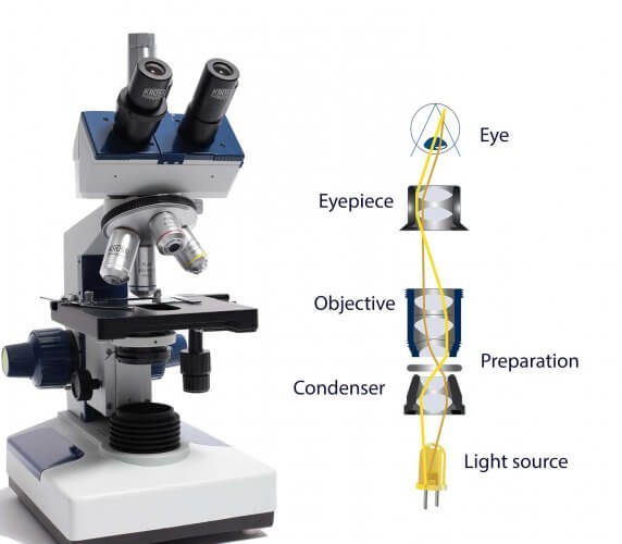
How do microscopes operate? - Krüss laboratory equipment
Compound Microscope: Definition, Diagram, Parts, Uses, Working ... - BYJUS Compound microscope is a type of optical microscope that is used for obtaining a high-resolution image. There are more than two lenses in a compound microscope. Learn about the working principle, parts and uses of a compound microscope along with a labeled diagram here.

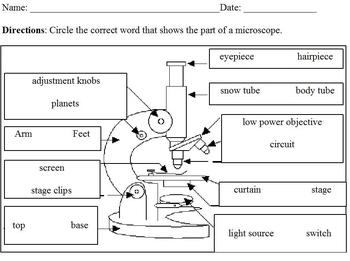
Label a Microscope Worksheet
Microscope Diagram - Label Diagram | Quizlet The bottom of the microscope, used for support. ocular lens. Eyepiece of a microscope. Diaphragm. Regulates the amount of light on the specimen. nosepiece of microscope. holds the objective lenses. objective lens. The lens on a light microscope that is closest to the stage.

Microscope Labeling Diagram | Quizlet
Welcome to Butler County Recorders Office Copy and paste this code into your website. Your Link …

Compound Microscope: Parts of Compound Microscope
Compound Microscope Parts, Functions, and Labeled Diagram Compound Microscope Parts, Functions, and Labeled Diagram Parts of a Compound Microscope Each part of the compound microscope serves its own unique function, with each being important to the function of the scope as a whole.
Label Microscope Parts - ClipArt Best
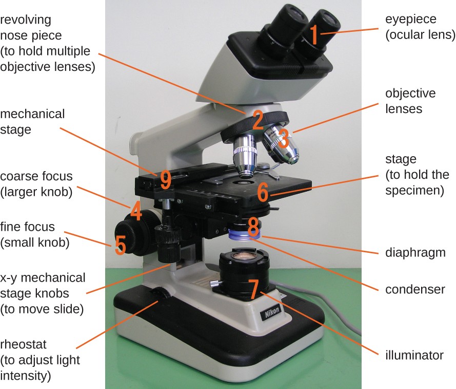
researchtweet.com › microscope-parts-labeledMicroscope, Microscope Parts, Labeled Diagram, and Functions Jan 19, 2022 · The liquid sample comes next. To minimise evaporation and protect the microscope lens from sample exposure, a small square of clear glass or plastic (a coverslip) is placed on top of the liquid. 1. Collect a clean microscope slide and a coverslip (a thin piece of plastic covering). Fill the centre of the microscope slide with a drop or two of ...
Glossary of terms used in microscopy – Quekett Microscopical Club
A Study of the Microscope and its Functions With a Labeled Diagram ... A Study of the Microscope and its Functions With a Labeled Diagram To better understand the structure and function of a microscope, we need to take a look at the labeled microscope diagrams of the compound and electron microscope. These diagrams clearly explain the functioning of the microscopes along with their respective parts.

microscope drawing with label - Clip Art Library
Parts of Stereo Microscope (Dissecting microscope) – labeled diagram ... Labeled part diagram of a stereo microscope Major structural parts of a stereo microscope. ... Apart from magnification, some eyepieces come with the label “WF” which defines that the eyepiece provides a wide field of view. This means that while viewing the specimen, the user will see wider areas compared to the field of view perceived ...

Labeled Microscope Diagram | Microscope parts, Science fair ...
depts.washington.edu › vurchinWelcome to Virtual Urchin - University of Washington Major update Apr 2021: All of the activities on the site are now mobile compatible !! Computers are still recommended, and tablets are preferable to phones: please read the Notes at the bottom of this page for details on the latest updates, mobile compatibility and general information about using this site.

Label the microscope — Science Learning Hub
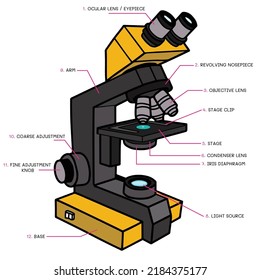
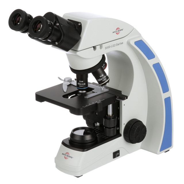
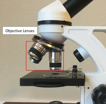
Microscope, Microscope Parts, Labeled Diagram, and Functions Jan 19, 2022 · Revolving Nosepiece or Turret: Turret is the part of the microscope that holds two or multiple objective lenses and helps to rotate objective lenses and also helps to easily change power. Objective Lenses: Three are 3 or 4 objective lenses on a microscope. The objective lenses almost always consist of 4x, 10x, 40x and 100x powers. The most common eyepiece …
Parts of a microscope with functions and labeled diagram
Simple Microscope - Diagram (Parts labelled), Principle, Formula and Uses Parts of a Simple Microscope A simple microscope consists of Optical parts Mechanical parts Labeled Diagram of simple microscope parts Optical parts The optical parts of a simple microscope include Lens Mirror Eyepiece Lens A simple microscope uses biconvex lens to magnify the image of a specimen under focus.

Compound Microscope Parts, Functions, and Labeled Diagram ...
› ternary-phase-diagramTernary Phase Diagram - an overview | ScienceDirect Topics A point on the diagram represents a composition that is specified in terms of mole fraction or weight fraction. The point, (0.3, 0.4, 0.3) is at the center of the small triangle in the diagram and is located by following the red diagonal 60° line at red 0.3 and the horizontal line at blue 0.4 or any combination of two of the coordinates (A, B, C).

How to draw compound of Microscope easily - step by step
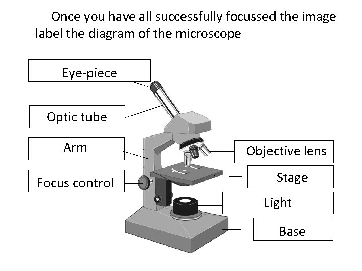
Label the microscope — Science Learning Hub Jun 08, 2018 · All microscopes share features in common. In this interactive, you can label the different parts of a microscope. Use this with the Microscope parts activity to help students identify and label the main parts of a microscope and then describe their functions.. Drag and drop the text labels onto the microscope diagram. If you want to redo an answer, click on the …

Compound Microscope Parts – Labeled Diagram and their ...
National Geographic Magazine National Geographic stories take you on a journey that’s always enlightening, often surprising, and unfailingly fascinating.

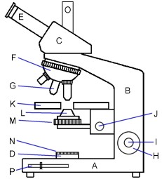
This is a common compound microscope. Label its parts from A ...
2001 anthrax attacks - Wikipedia The 2001 anthrax attacks, also known as Amerithrax (a portmanteau of "America" and "anthrax", from its FBI case name), occurred in the United States over the course of several weeks beginning on September 18, 2001, one week after the September 11 terrorist attacks.Letters containing anthrax spores were mailed to several news media offices and to Democratic …

Label the Microscope Diagram | Download Scientific Diagram
Compound Microscope Parts – Labeled Diagram and their … Major structural parts of a compound microscope. There are three major structural parts of a compound microscope. The head includes the upper part of the microscope, which houses the most critical optical components, and the eyepiece tube of the microscope.; The base acts as the foundation of microscopes and houses the illuminator.; The arm connects between the base …
Microscope Biology - 2022
Microscope Types (with labeled diagrams) and Functions Simple microscope labeled diagram Simple microscope functions It is used in industrial applications like: Watchmakers to assemble watches Cloth industry to count the number of threads or fibers in a cloth Jewelers to examine the finer parts of jewelry Miniature artists to examine and build their work Also used to inspect finer details on products

Microscope parts 3D learning for Android - APK Download
Family Tree Stock Photos, Pictures & Royalty-Free Images A set of genetics icons that include editable strokes or outlines using the EPS vector file. The icons include families, DNA, Genes, genetic testing concepts, cells, science and biology, microscope, geneticists, family tree, biologist using microscope, petri dish, human biology, human identity and other related icons.
Modified Science Diagram; Label Parts of a Microscope; Special Education
Microscope Using a microscope I have developed my

Parts of a Microscope - SmartSchool Systems

5 Important Types of Microscopes used in Biology (With Diagram)
Instruments of Microscopy | Microbiology | | Course Hero
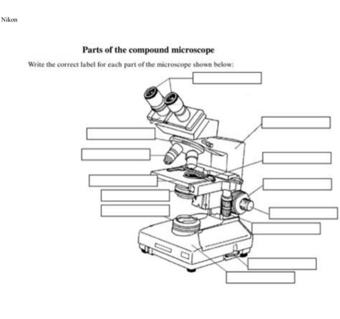
Solved Nikon Parts of the compound microscope Write the ...

The Microscope
Microscope, Microscope Parts, Labeled Diagram, and Functions

Compound Microscope- Definition, Labeled Diagram, Principle ...
40,226 Light microscope Images, Stock Photos & Vectors ...

Light Microscope- Definition, Principle, Types, Parts ...

A schematic diagram for the microscope-based label-free ...

Label the microscope — Science Learning Hub
Microscope labeled diagram
Compound Microscope Parts, Functions, and Labeled Diagram ...

✓ microscope view free vector eps, cdr, ai, svg vector ...

File:Labelledmicroscope.gif - Wikimedia Commons

Parts of a Compound Microscope and Their Functions

Lable the microscope worksheet
Free Microscope Drawing, Download Free Microscope Drawing png ...
16 Parts of a Compound Microscope: Diagrams and Video ...

Labeling the Parts of the Microscope | Microscope World Resources
Post a Comment for "41 diagram of a microscope and label"