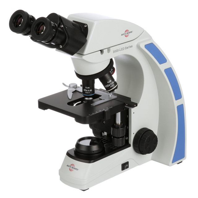
41 microscope no labels
Amazing 27 Things Under The Microscope With Diagrams Amazing 27 Things Under The Microscope With Diagrams February 20, 2022 by Anupama Sapkota Note: Each image source is given below in this post of respective subheadings. Table of Contents 1. Amoeba under the microscope Direct observation Observation after staining 2. Algae under the microscope Chlorophyta Chromophyta Cryptophyta Rhodophyta Microscope Notes - Northern Arizona University Microscope Drawings. When drawing what you see under the microscope, follow the format shown below. It is important to include a figure label and a subject title above the image. The species name (and common name if there is one) and the magnification at which you were viewing the object should be written below the image.
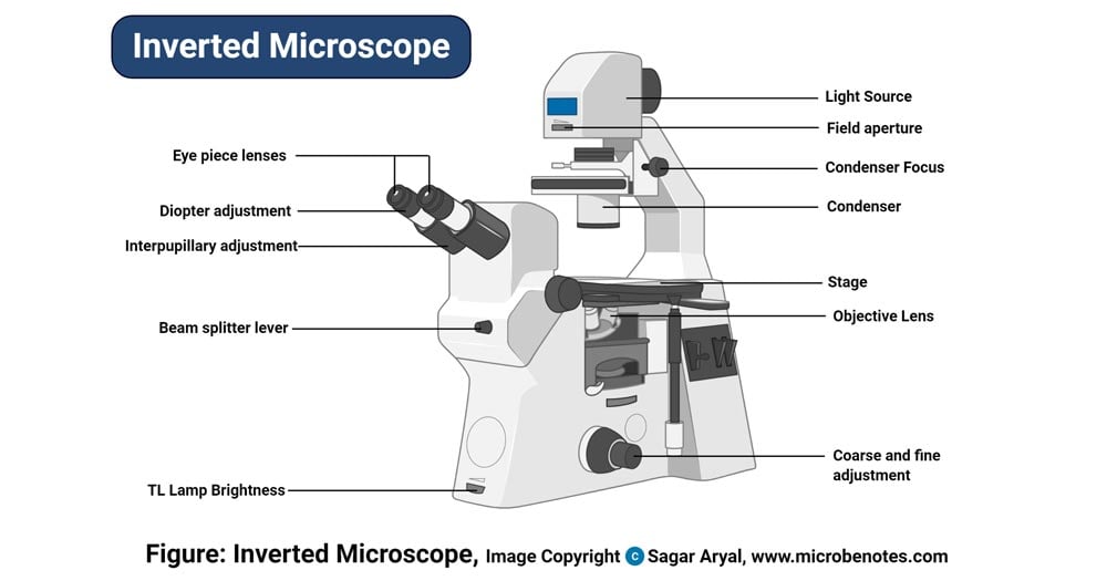
Parts of a microscope with functions and labeled diagram Microscope Definition Microscopes are instruments that are used in science laboratories to visualize very minute objects such as cells, and microorganisms, giving a contrasting image that is magnified. Microscopes are made up of lenses for magnification, each with its own magnification powers.

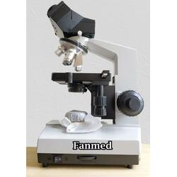
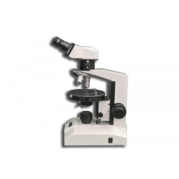
Microscope no labels
Microscope Parts and Functions A standard microscope has three, four, or five objective lenses that range in power from 4X to 100X. When focusing the microscope, be careful that the objective lens doesn't touch the slide, as it could break the slide and destroy the specimen. Specimen or slide: The specimen is the object being examined. enchantedlearning.com Moved Permanently. The document has moved here. Compound Microscope Parts - Labeled Diagram and their Functions - Rs ... The eyepiece (or ocular lens) is the lens part at the top of a microscope that the viewer looks through. The standard eyepiece has a magnification of 10x. You may exchange with an optional eyepiece ranging from 5x - 30x. [In this figure] The structure inside an eyepiece. The current design of the eyepiece is no longer a single convex lens.
Microscope no labels. Microscope Labeling Game - PurposeGames.com An unregistered player played the game 5 minutes ago About this Quiz This is an online quiz called Microscope Labeling Game There is a printable worksheet available for download here so you can take the quiz with pen and paper. This quiz has tags. Click on the tags below to find other quizzes on the same subject. Science microsope A Study of the Microscope and its Functions With a Labeled Diagram Here, unlabeled microscope diagrams have been provided for your perusal, which will help you practice and test your understanding of the instrument. Types of Microscopes Depending on the source of illumination, microscopes can be divided into two categories. They are: Microscope, Microscope Parts, Labeled Diagram, and Functions Microscopes magnify or enlarge small objects such as cells, microbes, bacteria, viruses, microorganisms etc. at a viewable scale for examination and analysis. Microscopes consist of one or more magnification lenses to enlarge the image of the microscopic objects placed in the focal plane. Microscope: Label the Parts Flashcards | Quizlet Match each part to the picture! Learn with flashcards, games, and more — for free.
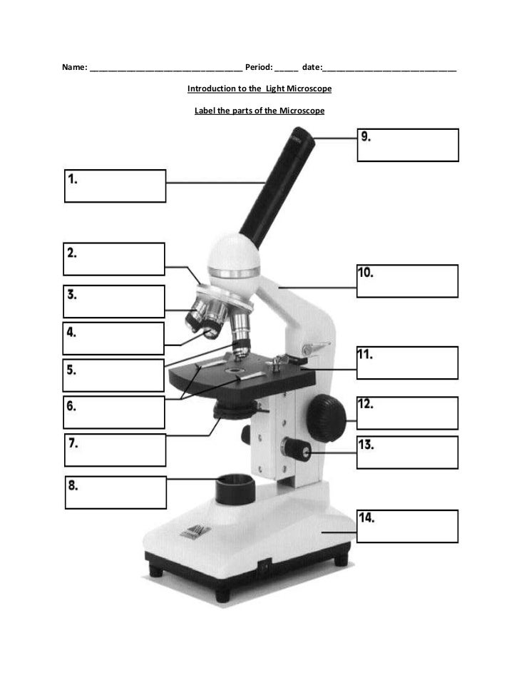
Specimen Labeling Requirements - Department of Pathology Blood Bank sample labels (crossmatch or type & hold) must be handwritten. Addressograph labels must not be placed on crossmatch specimens. A specimen is incompletely labeled if some of the required information is missing: If the name and the medical record number are missing, the specimen will be considered unlabeled and handled as such. Compound Microscope - Diagram (Parts labelled), Principle and Uses What are the 13 parts of a microscope? 1. Eyepiece 2. Eyepiece Tube 3. Objective Lens 4. Stage 5. Stage Clips 6. Nosepiece 7. Fine and Coarse Focus knobs 8. Illuminator 9. Aperture 10. Iris Diaphragm 11. Condenser 12. Condenser Focus Knob 13. The Rack stop Q 5. What are the 11 parts of a compound microscope? Amazon.com: Microscopes - Binoculars & Scopes: Electronics: USB ... 100X-2000X Microscopes for Kids Students Adults, with Microscope Slides Set, Phone Adapter, Powerful Biological Microscopes for School Laboratory Home Education. 4.4 out of 5 stars 1,651-33% $99.99 $ 99. 99 $150.00 $150.00. Get it as soon as Fri, Jul 15. FREE Shipping by Amazon. Microscope Magnification: Explained - Microscope Clarity To calculate the magnification on a microscope multiply the magnification power of the eyepiece you are using by the objective currently in position. Magnification = Eyepiece Magnification X Objective Magnification Microscopes magnify or enlarge the image under inspection and enables the human eye to see things we would never be able to see.
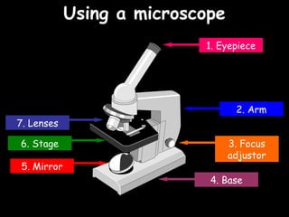
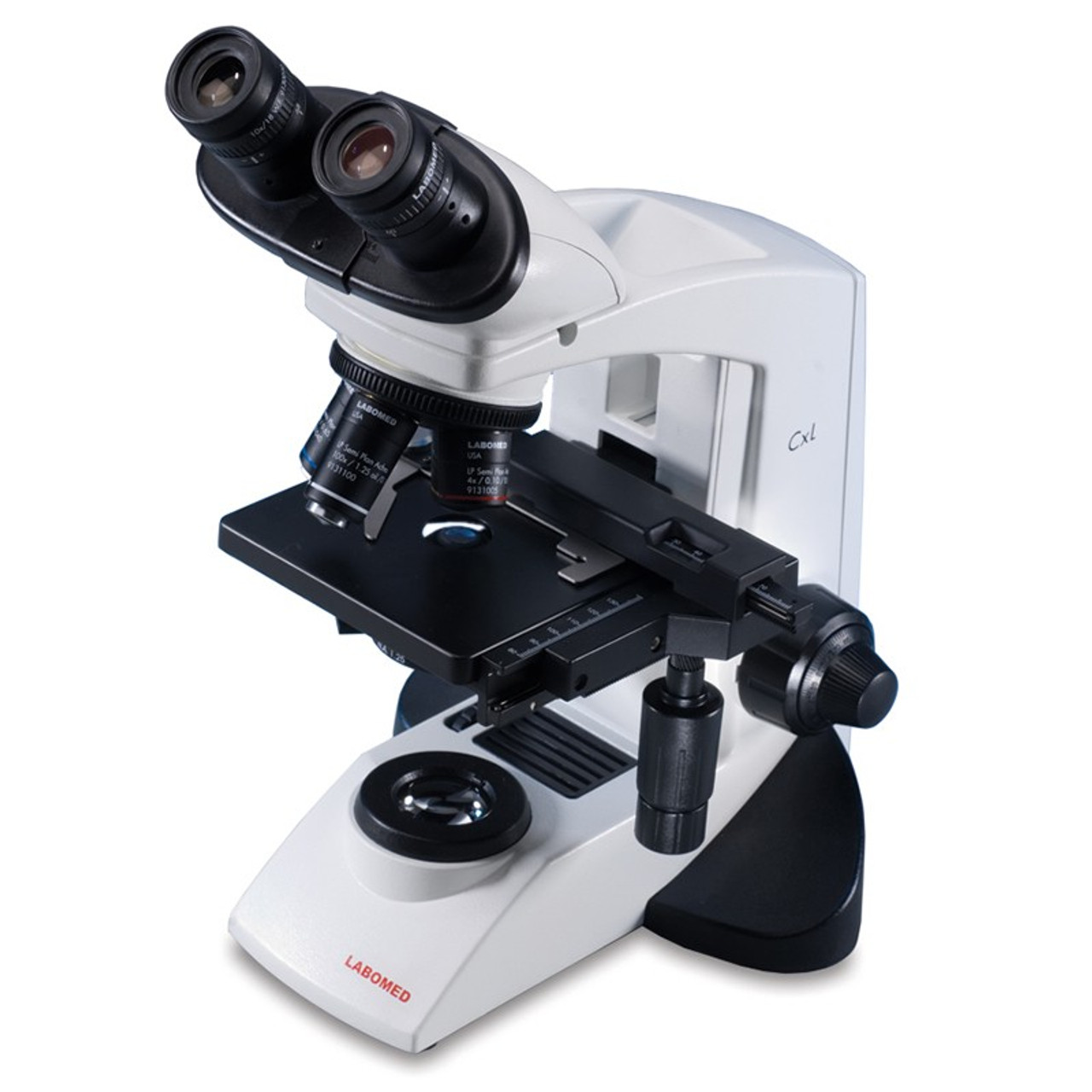
What is a Compound Microscope? - BYJUS The compound microscope is mainly used for studying the structural details of cell, tissue, or sections of organs. The parts of a compound microscope can be classified into two: Non-optical parts Optical parts Non-optical parts Base The base is also known as the foot which is either U or horseshoe-shaped. Label the microscope — Science Learning Hub All microscopes share features in common. In this interactive, you can label the different parts of a microscope. Use this with the Microscope parts activity to help students identify and label the main parts of a microscope and then describe their functions. Drag and drop the text labels onto the microscope diagram. Microscope.com - Affordable microscopes for everyday use The Microscope Experts. Founded by a high school biology teacher in 1998, Microscope.com is now the largest and most trusted online retailer of professional quality, affordable microscopes. Our success is based on an unerring commitment to expert and friendly customer service. We are here to help you find the best microscope for your needs at ... Virtual Microscope | NCBioNetwork.org Learning Objectives. Upon completion of this exercise, you will be able to: List the basic components of a typical microscope. Describe the use of lens power and eyepiece powers. Calculate the magnification of a microscope based on the selected lens. Discuss the care of an use of a typical microscope.
Labeling the Parts of the Microscope | Microscope World Resources
Free Microscope Worksheets for Simple Science Fun for Your Students The first worksheet labels the different parts of a microscope, including the base, slide holder, and condenser. I f you have a microscope, compare and contrast this worksheet to it. Also, your kids can color this microscope diagram in and read the words to each part of the microscope.

Metalurgi Microscop AmScope Perlengkapan 40X 1000X EPI ...
The Virtual Microscope - An online Interactive look at Microscopy The virtual microscope allows people all over the world to study specimens when ever and however they want. Unlike prepared slides or photographs, virtual images allow viewers to target a certain spot of a specimen for detailed study and provides the tools to manipulate the image to see what it looks like from a different angle and more.

microscope | Types, Parts, History, Diagram, & Facts | Britannica
Labeling the Parts of the Microscope Labeling the Parts of the Microscope This activity has been designed for use in homes and schools. Each microscope layout (both blank and the version with answers) are available as PDF downloads. You can view a more in-depth review of each part of the microscope here. Download the Label the Parts of the Microscope PDF printable version here.
Compound Microscope Parts, Functions, and Labeled Diagram ...
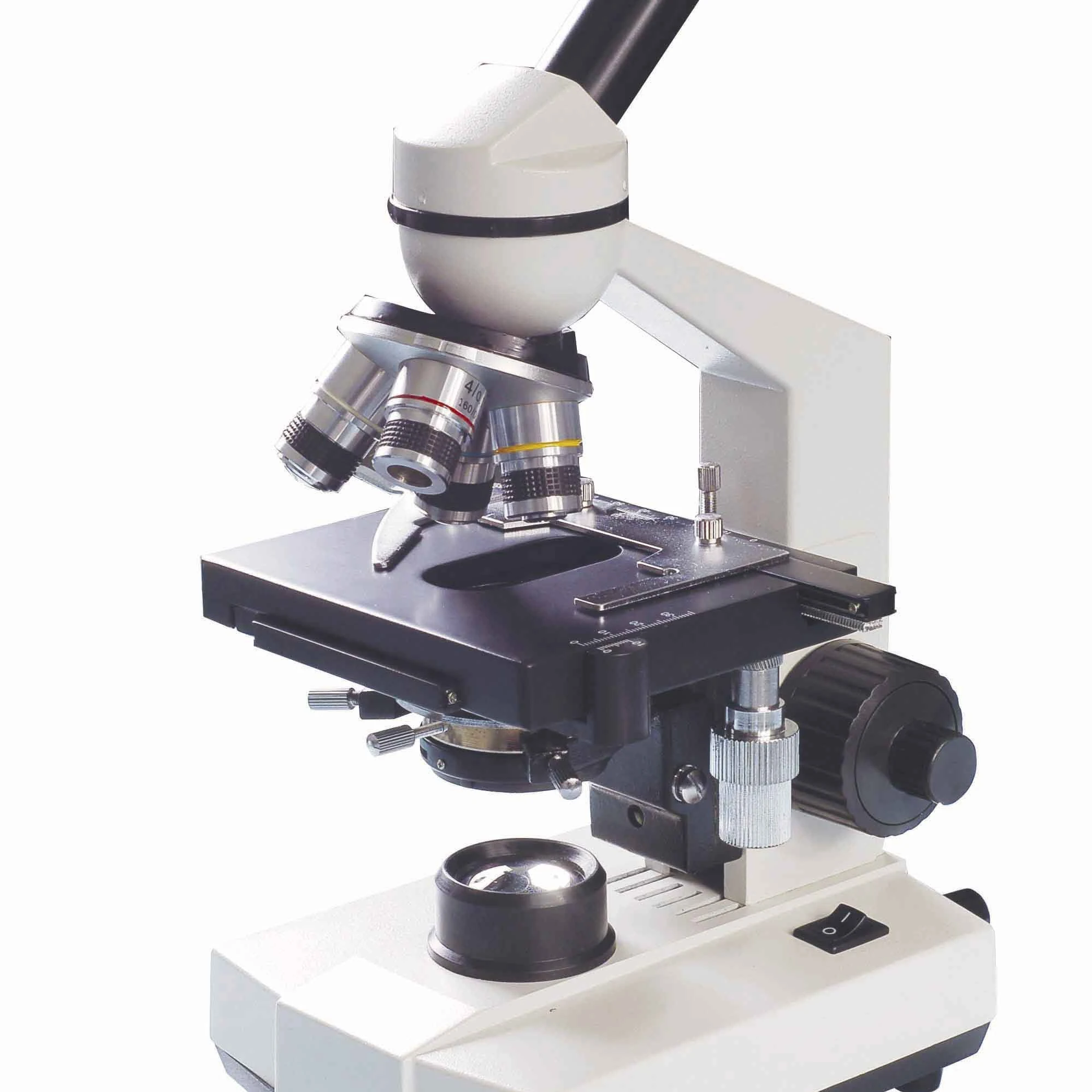
Parts of the Microscope with Labeling (also Free Printouts) Parts of the Microscope with Labeling (also Free Printouts) A microscope is one of the invaluable tools in the laboratory setting. It is used to observe things that cannot be seen by the naked eye. Table of Contents 1. Eyepiece 2. Body tube/Head 3. Turret/Nose piece 4. Objective lenses 5. Knobs (fine and coarse) 6. Stage and stage clips 7. Aperture

AmScope 40-2000x Digital Binocular Compound Microscope + Built-in 3MP USB Camera 670541565476 | eBay
Looking at the Structure of Cells in the Microscope A typical animal cell is 10-20 μm in diameter, which is about one-fifth the size of the smallest particle visible to the naked eye. It was not until good light microscopes became available in the early part of the nineteenth century that all plant and animal tissues were discovered to be aggregates of individual cells. This discovery, proposed as the cell doctrine by Schleiden and Schwann ...

Label the parts and functions of the microscope no. 1-16 ...
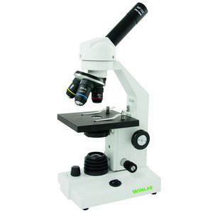
Simple Microscope - Parts, Functions, Diagram and Labelling A compound microscope is also called a bright field microscope. It can provide magnification by up to 1,000 times. Stereo microscope/dissecting microscope - It can magnify objects by up to 300 times. It is used to visualize opaque objects that cannot be visualized using a compound microscope.
Jual Mikroskop Microscope Biological Monokuler XSP- 12 Series ...
Simple Microscope - Diagram (Parts labelled), Principle, Formula and Uses The working principle of a simple microscope is that when a lens is held close to the eye, a virtual, magnified and erect image of a specimen is formed at the least possible distance from which a human eye can discern objects clearly. Magnification formula The magnification power of a simple microscope is expressed as: M = 1 + D/F Where

Bau des Mikroskops by frau_sommarkind on Genially
Troubleshooting Microscope Lighting and Light Bulb Issues Make sure the microscope is plugged in and switched on. Many microscopes have a rheostat control (either a knob or a small flat dial built into the microscope base). Make sure the rheostat control is turned up to the brightest level. Is the microscope field iris opened up? The microscope field iris is part of the microscope condenser.

Nanolive 3D Cell Explorer CX-A | Microscopes & Image Analysis ...
Microscope not recognized or missing - Dino-Lite Right-click on Computer, My Computer, or This PC on your desktop or start menu, then select Manage In the computer management window, select Device Manager In the Device Manager window, open the Imaging Devices category, or, if that category is not visible, open the Other Devices category.

Parts of a microscope with functions and labeled diagram
Solved Ashley lost the labels to her microscope slides and | Chegg.com Question: Ashley lost the labels to her microscope slides and can no longer tell the difference between the two. Explain how she should distinguish a slide of the epithelium lining the esophagus from a slide of the epithelium lining the small intestine. This problem has been solved!
PROPOSAL TUGAS AKHIR – SB 091351 PENGARUH PEMBERIAN MIKORIZA ...
Anatomy of the Microscope - Microscope Stages | Olympus LS A simple (commonly termed "plain") microscope stage is illustrated on the left in Figure 2. This stage contains an opening to admit light from the condenser, several mounting holes for a mechanical stage, and two clips that secure the specimen slide in place for observation under increasing magnification (changing of objectives) and for photomicrography.

Pin on Products
Compound Microscope Parts - Labeled Diagram and their Functions - Rs ... The eyepiece (or ocular lens) is the lens part at the top of a microscope that the viewer looks through. The standard eyepiece has a magnification of 10x. You may exchange with an optional eyepiece ranging from 5x - 30x. [In this figure] The structure inside an eyepiece. The current design of the eyepiece is no longer a single convex lens.
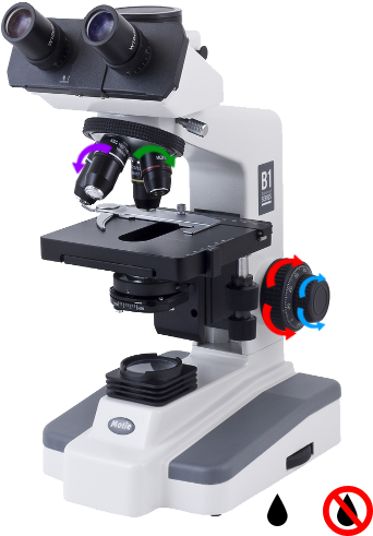
Operating instructions
enchantedlearning.com Moved Permanently. The document has moved here.

solomark hm900 upright monocular microscope
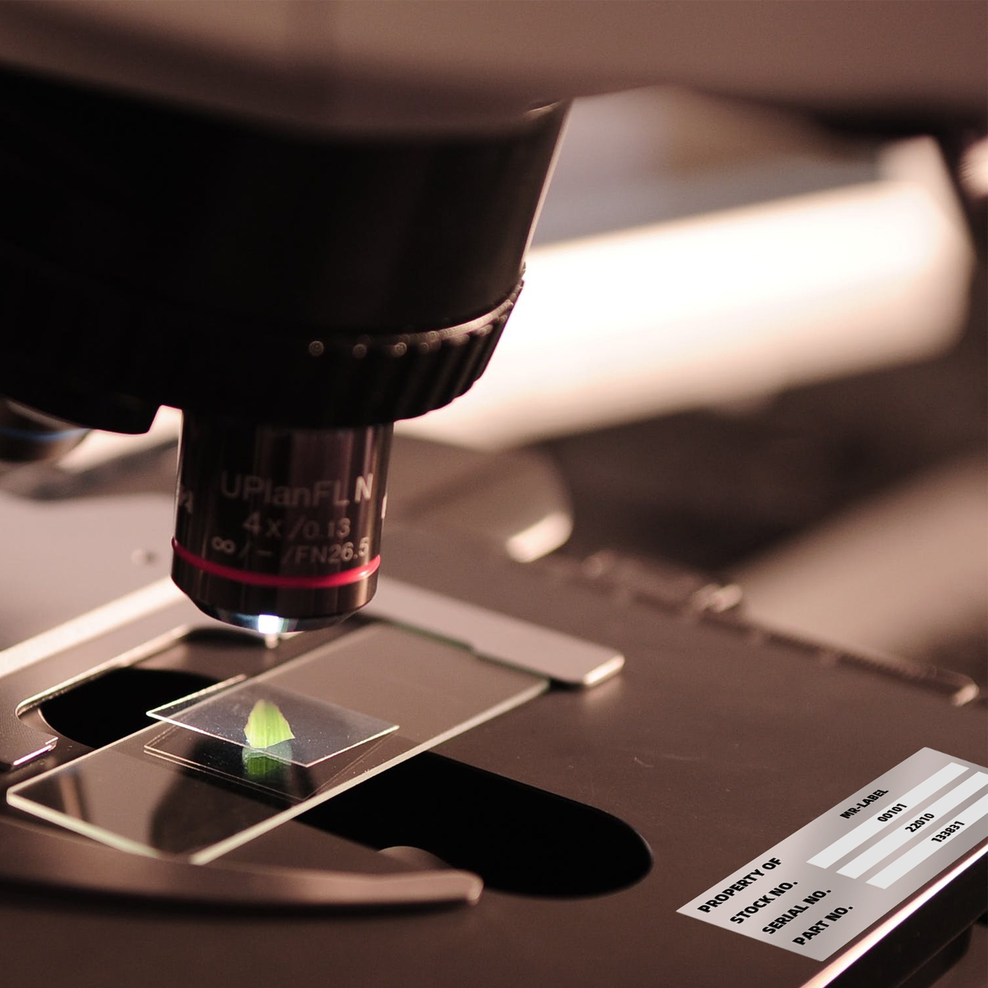
Microscope Parts and Functions A standard microscope has three, four, or five objective lenses that range in power from 4X to 100X. When focusing the microscope, be careful that the objective lens doesn't touch the slide, as it could break the slide and destroy the specimen. Specimen or slide: The specimen is the object being examined.
How to Use the Microscope

Step Stereo Microscope Archives - Lab Equipment|Chemistry Lab ...

Label the microscope — Science Learning Hub

Biology 11/09/18 | Ms Chenwen's Home Room

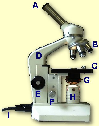
This is a common compound microscope. Label its parts from A ...

Wolfe® Cordless Advanced LED Binocular Microscope

TYPES OF LIGHT MICROSCOPE: 1. SIMPLE... - Microbiologists_2 ...
ML9200L

microscope vector sketch 7314112 Vector Art at Vecteezy

Cole-Parmer Economical Compound Microscope, 115 VAC from Cole ...
Download Microscope With Labels PNG Image with No Background ...
Mr-Label 63.5 x 29.6 mm Silver Asset Stickers – Self-Adhesive ...

electron microscope to label - Clip Art Library
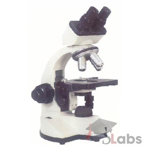
Co-axial-Binocular-Microscope type-2 - Scholars Labs

Laboratory Equipment – My Blog
Mikroskop Xsp-104 Biologi Pelajar Monokular - Buy Bermata Biological Microscope,Salah Satu Tujuan Mikroskop,Siswa Biological Microscope Product on ...
Biology label part of microscope
Microscope
7 a cells
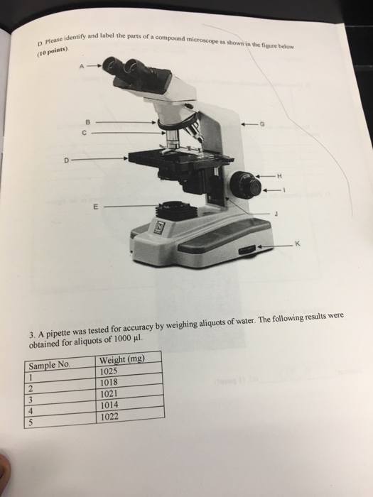
Solved Identify and label the parts of a compound microscope ...
Parts of a microscope with functions and labeled diagram
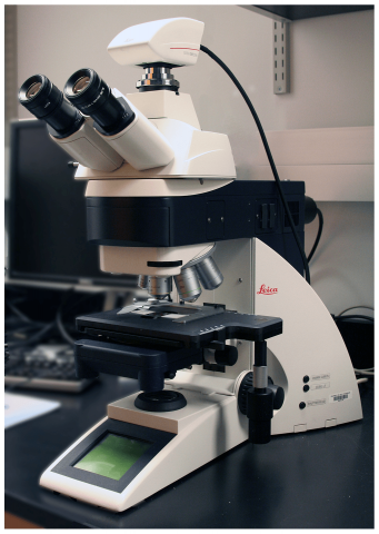
Leica DM 5500B Compound Microscope with Leica DFC290 Color ...
Compound Microscope Parts, Functions, and Labeled Diagram ...

7" LCD Digital Microscope W/32GB, 50x-2000x Dual Lens Cell ...
Windaus Microscope HPM 100 LED

Metallurgical Microscope Market Is Expected To Be Valued At ...
Untitled Document
Post a Comment for "41 microscope no labels"